IMAGE

Figure 3

ID
ZDB-IMAGE-221118-52
Source
Figures for Rehman et al., 2022
Image
Figure Caption

Figure 3

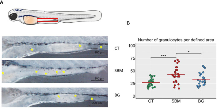
Localization of granulocytes in the intestine of zebrafish larvae. (A) Representative images of the intestine region of zebrafish larvae stained with Sudan Black to reveal the presence of granulocytes, yellow arrows indicate the granulocytes in the intestine region. (B) Quantification of Sudan-Black+ cells in the intestine region of zebrafish larvae. *** p < 0.001 and * p < 0.05. n = 24-25 per group. Control - CT, soybean - SBM and β-glucan – BG. Scale bar = 200 μm.

Acknowledgments
This image is the copyrighted work of the attributed author or publisher, and ZFIN has permission only to display this image to its users. Additional permissions should be obtained from the applicable author or publisher of the image. Full text @ Front Immunol