
|
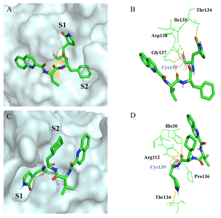
Fig. 7
Molecular docking of compound 10d in the GI.1 and GII.4 norovirus 3CL protease. (A) The binding mode of 10d at the substrate-binding site of the GI.1 norovirus 3CLpro (PDB code: 3UR9). The GI.1 norovirus 3CLpro was shown as molecular surface and 10d was shown by green sticks. (B) Interactions of 10d with the surrounding residues. Residues are shown as light green lines, and H-bonds are represented by yellow dashed lines. (C) The binding mode of 10d at the substrate-binding site of the GII.4 norovirus 3CLpro (PDB code: 6NIR). (D) Interactions of 10d with the surrounding residues. The Schrödinger program (Maestro covalent docking) was used for calculations and PyMOL program for visualizations.