
|
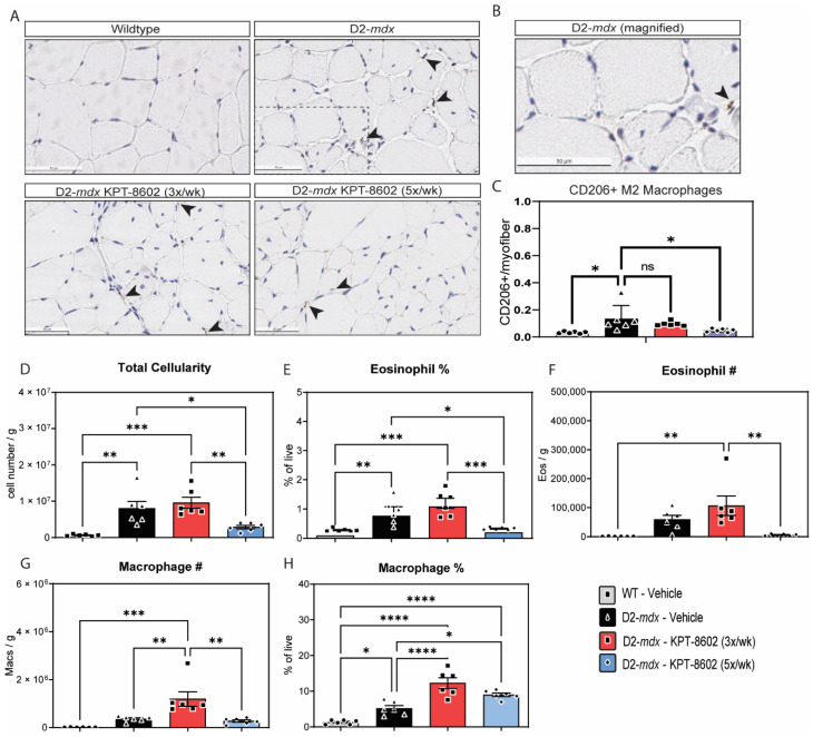
Figure 3
KPT−8602 treatment shifts D2-mdx muscles to a pro-regenerative, M2 macrophage profile. (A) Immunostaining of CD206-positive TA muscles from WT-vehicle, D2-mdx vehicle, D2-mdx KPT−8602 3x/week, and D2-mdx 5x/week experimental mouse cohorts. Arrows indicate CD206 stained macrophages. Boxed area magnified in B. Scale bar = 50 µm; (B) Magnified inset of a CD206+ macrophage from the D2-mdx sample; (C) Quantification of the total number of CD206+ M2 macrophages normalized to the total number of myofibers per area quantified to a minimum of 500 myofibers. All data is presented as mean ± SEM, n = 6; * p < 0.05, ** p < 0.01, *** p < 0.001, **** p < 0.0001; one-way ANOVA with Tukey’s HSD test); (D) Total cellularity measured in quadricep muscle cell preparations from the 4 experimental cohorts: WT vehicle (grey), D2-mdx vehicle (black), D2-mdx 3x/week (red); D2-mdx 5x/week (blue); (E,F) graphs showing the percentage of eosinophils and the total eosinophil number/gram muscle wet weight; (G,H) Graphs showing the percentage of macrophages and the total macrophage number/gram muscle wet weight.