
Image
|
Figure Caption
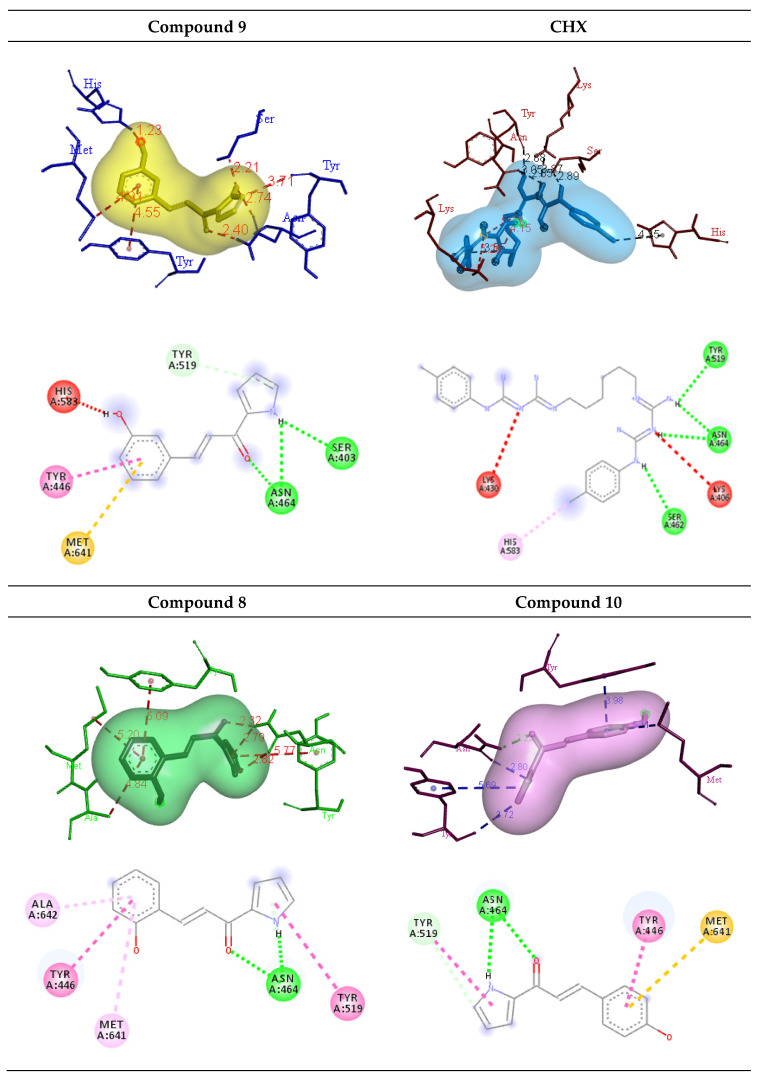
Fig. 3
3D and 2D diagrams of the binding interaction of hydroxylated compounds 8, 9, and 10 (green line: H bond; light green line; carbon H bond/π-donor H bond; red line: donor-donor; yellow line: π-sulphur; pink line: π-π stacked/π-π T-shaped; light pink line: π-alkyl) and CHX (green line: H bond; red line: positive-positive; light purple line: π -alkyl) with the amino acid residues on the active site of the PBP2a receptor.
Acknowledgments
This image is the copyrighted work of the attributed author or publisher, and
ZFIN has permission only to display this image to its users.
Additional permissions should be obtained from the applicable author or publisher of the image.
Full text @ Molecules