
|
Fig. 1
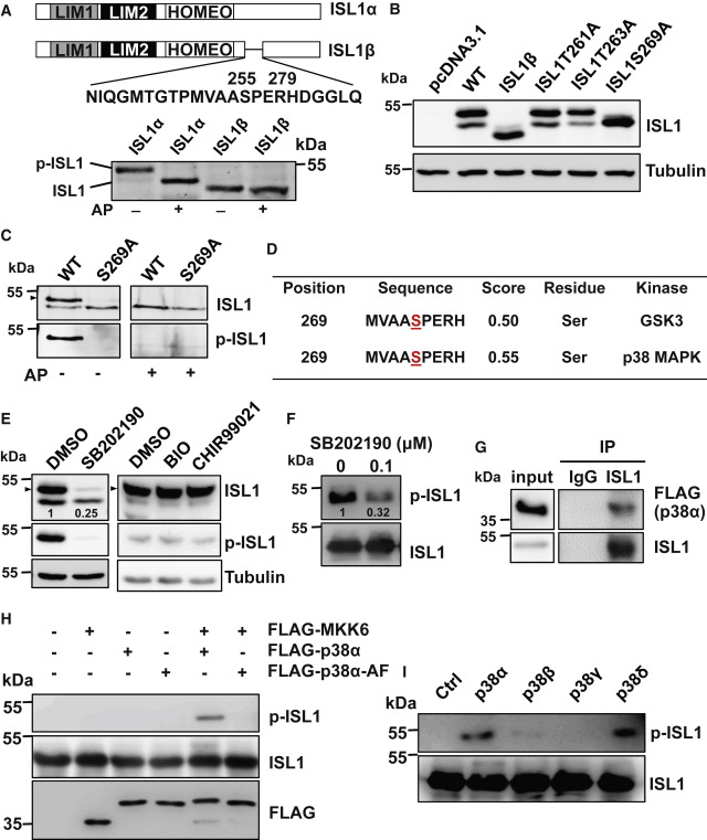
Figure 1. ISL1 is phosphorylated by p38 on serine 269 (A) Schematic representation of ISL1α and ISL1β (top panel). Alkaline phosphatase (AP) treatment of extracts from cells expressing either ISL1α or ISL1β, showing that the β form is not phosphorylated. (B) Western blot (WB) analysis of extracts of cells expressing ISL1α harboring different mutations in the serine or threonine residues within the 256–278 region. Tubulin served as loading control. (C) AP treatment of extracts from cells expressing either WT ISL1α or ISL1αS269A, showing that ISL1αS269A mutant is not phosphorylated. Antibody raised against synthetic ISL1 phospho-S269-peptide detected phosphorylated ISL1 only in extracts overexpressing ISL1 but not the S269A mutant. Arrowheads in this and the following figure panels indicate phosphorylated ISL1. (D) Results of predictions of kinase-specific protein phosphorylation sites using NetPhosK1.0. The potentially modified residues are underlined. (E) WB analysis of ISL1 overexpressing HEK293T cells treated either with DMSO, 50 μM p38 inhibitor SB202190, or with the GSK3 inhibitors, 2.5 μM BIO and 5 μM CHIR-99021 for 16 h using ISL1 and phospho-ISL1 antibody. (F) In vitro kinase assay using recombinant ISL1 incubated with total protein extracts from cells treated with or without SB202190. (G) Co-immunoprecipitation of protein extracts of HEK293T cells overexpressing ISL1 and FLAG-p38α using ISL1 antibody followed by WB analysis with FLAG and ISL1 antibody. (H) In vitro kinase assay with recombinant ISL1 incubated with combinations of immunopurified MKK6, p38α, and kinase-deficient p38α (p38α-AF) proteins. FLAG-tagged constructs for these kinases were overexpressed alone or in combinations and immunoprecipitated using FLAG antibody. (I) In vitro kinase assay with recombinant ISL1 incubated with immunopurified MKK6 together with different p38 MAPK isoforms.