
|
Fig. 1
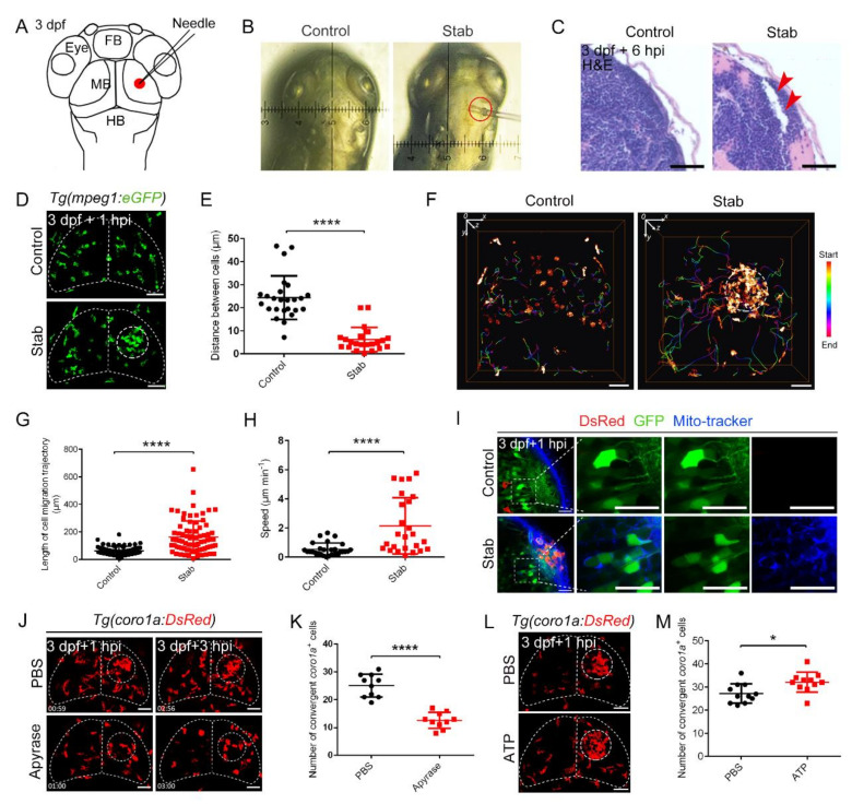
Predominant involvement of macrophages in response to traumatic brain injury caused by stabbing in zebrafish. (A) Stab model. The red dot marks the lesion core (LC). FB, forebrain; MB, midbrain; HB, hindbrain. (B) An actual operation diagram of the stab. The horizontal width of damage was about 130 μm. (C) A paraffin section was stained with hematoxylin and eosin (H&E) staining. The red arrowheads indicate the brain tissue loss. Scale bar, 20 µm. (D) The accumulation of macrophages in Tg(mpeg1:GFP) at 1 hpi. The white dotted line shows the major area of macrophage aggregation. Scale bar, 50 µm. (E) Statistical analysis of the distance between coro1a+ cells in LC. Control, 24.35 ± 1.89 µm n = 25; Stab, 6.14 ± 1.12 µm n = 22. (F) Spatial and temporal migration of macrophages. Scale bar, 50 µm. (G) Length statistics for macrophage migration trajectories. Control, 62.14 ± 3.881 µm, n = 71; Stab, 161.7 ± 13.42 µm, n = 78. (H) Statistical analysis of the movement speed of coro1a-DsRed+ cells after injury. Control, 0.5309 ± 0.08210 µm min−1 n = 28; Stab, 2.148 ± 0.3875 µm min−1 n = 25. (I) MitoTracker signals were detected in the damage sites. The right three panels are the enlarged views of the boxed regions in the left panel. Scale bar, 20 µm. (J) Aggregation of macrophages after apyrase injection in Tg(coro1a:DsRed) embryos at 1 hpi and 3 hpi. Scale bar, 50 µm. (K) Statistical number of coro1a+ cells after injury at 1 hpi. PBS, 25.10 ± 1.303 n = 10; apyrase, 12.60 ± 0.9092 n = 10. (L) Observation of macrophage aggregation after ATP injection at 1 hpi. Scale bar, 50 µm. (M) Statistical number of aggregated coro1a+ cells in (L). PBS, 27.18 ± 1.278 n = 11; ATP, 32.09 ± 1.317 n = 11. (Data are shown as mean ± SEM. *, p < 0.05; ****, p < 0.0001.)