
Image
|
Figure Caption
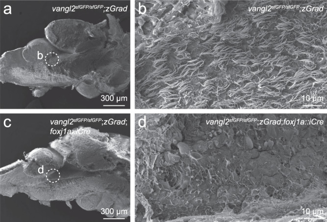
Fig. 7
FoxJ1a-lineage specific degradation of sfGFP-Vangl2 results in ependymal cell cilia defects.
Scanning electron micrographs of the rhombencephalic ventricle in brains dissected from 7-8 month old vangl2sfGFP/sfGFP;Tg(βactin2::loxP-mCherry-STOP-loxP-zGrad) control (a, b; n = 8) and vangl2sfGFP/sfGFP;Tg(βactin2::loxP-mCherry-STOP-loxP-zGrad);Tg(foxj1a::iCre) mutant (c, d; n = 8) zebrafish. Dashed circles in a, c indicate the area of high magnification images in b, d.
Figure Data
Acknowledgments
This image is the copyrighted work of the attributed author or publisher, and
ZFIN has permission only to display this image to its users.
Additional permissions should be obtained from the applicable author or publisher of the image.
Full text @ Nat. Commun.