IMAGE

Figure 3

ID
ZDB-IMAGE-220920-31
Source
Figures for Meyer-Miner et al., 2022
Image
Figure Caption

Figure 3

Re-introduction of Katnb1 in foxj1a-positive cell lineages suppresses scoliosis

(A) Schematic of Tg(foxj1a::katnb1) construct.

(B–E) SEM images of the rhombencephalic ventricle of 3 month old katnb1mh102/mh102 (B & C; N = 4, n = 11) and Tg(foxj1a::katnb1); katnb1mh102/mh102 (D & E; N = 4, n = 10) fish. (B, D) Scale bar, 20 μm. Square indicates higher magnification location. (C, E) Scale bar, 5 μm.

(F–I) Representative microCT projections of 3-month-old
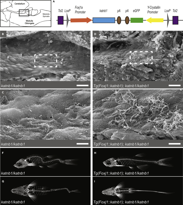
 katnb1mh102/mh102 (F & G; N = 17, n = 68) and Tg(foxj1a::katnb1); katnb1mh102/mh102 (H & I; N = 8, n = 73) mutant zebrafish in sagittal (F & H) and coronal (G & I) planes.

Figure Data
Acknowledgments
This image is the copyrighted work of the attributed author or publisher, and ZFIN has permission only to display this image to its users. Additional permissions should be obtained from the applicable author or publisher of the image. Full text @ iScience