
|
Fig. 8
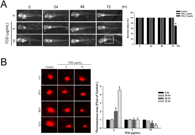
Fig. 8. Trichodermin inhibits tumor growth in a zebrafish xenograft model (A) TCD at the indicated concentrations and time had no observable toxicity effect at 72 hpi. *P < 0.05 compared with the control. (B) oral cancer HSC-3 cells labeled with a red fluorescent dye (CM‐DiI) were injected into 48 hpf normal zebrafish yolk sacs. The intensity of red fluorescence is proportional to the tumor size. Oral cancer xenograft zebrafish treated with 3 or 10 μg/mL TCD and observed at 24 and 48 hpi. Quantitative analysis of the tumor cell proliferation with or without TCD treatment. * *P < 0.01 compared with the control; *P < 0.05 compared with control cells treated with CPZ. Data represent means ± SD. hpf: hours post-fertilization; hpi: hours post-treatment or post-injection.