
|
Fig. 7
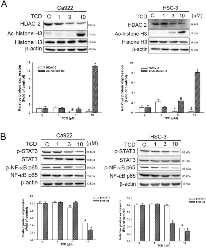
Fig. 7. Trichodermin decreases the HDCA-2-mediated signaling pathway in human OSCC cells. (A) OSCC cells (Ca922 and HSC-3 cells) were treated with the indicated concentrations of TCD, after which the expression levels of histone deacetylase 2 (HDAC-2), acetylated histone H3 (Ac-histone H3), histone H3 and β-actin were assessed by western blot analysis. (B) Levels of phospho-STAT3 (p-STAT3) and NF-κB (p- NF-κB) proteins were analyzed by Western blot assays. β-actin was used as an endogenous reference. Histograms represent the statistical analysis of the relative expression levels of phospho-STAT3 (p-STAT3) and NF-κB (p-NF-κB) proteins. Data represent means ± S.E.M. *P < 0.05, #P < 0.01 compared with the control.