
|
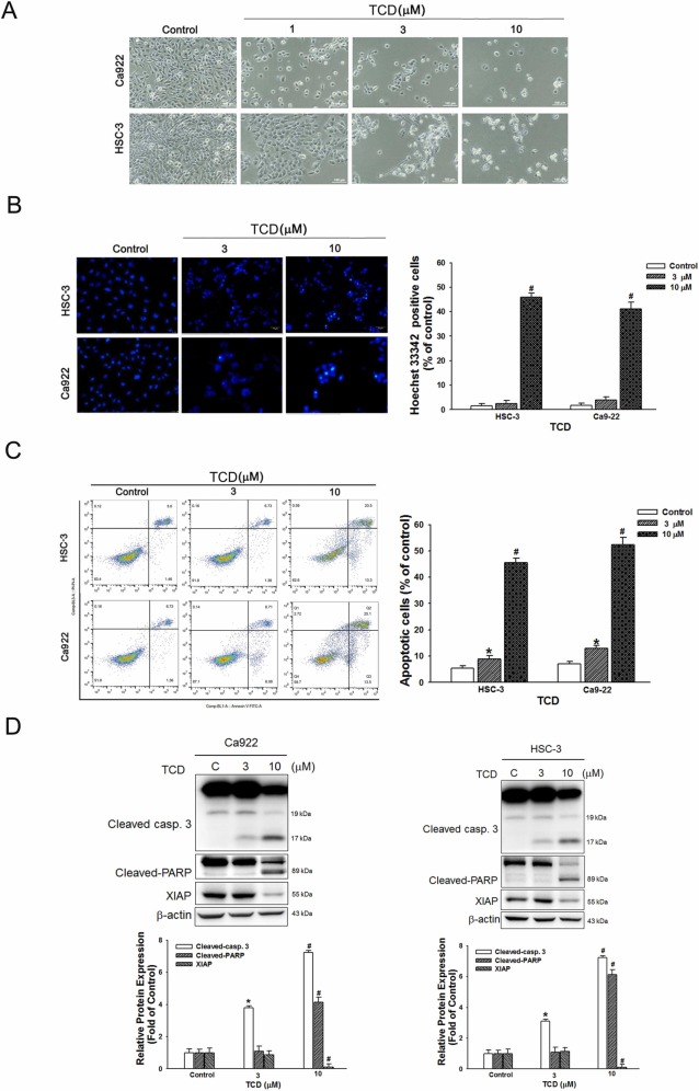
Fig. 4 Fig. 4. Trichodermin induces caspase-mediated apoptosis in human OSCC cells. (A) OSCC cells (Ca922 and HSC-3 cells) were treated with or without TCD for 24 h, after which apoptosis was monitored by phase contrast microscopy. Magnification, 20x. (B) Hoechst 33342 staining analysis of the apoptotic cell population in OSCC cells (Ca922 and HSC-3 cells) after exposure to 0, 3 or 10 µM TCD for 24 h. (C) Analysis of apoptosis was observed in TCD-treated OSCC cells (Ca922 and HSC-3 cells) for 24 h using Annexin V-FITC/PI staining with flow cytometry. Quantification analysis of the percentage of apoptotic cells is shown. (D) Cells were treated with TCD (0, 3 or 10 μM) for 24 h, after which the expression of apoptosis-associated proteins was determined by Western blots. β-actin was used as an endogenous reference. Histograms represent statistical analysis of the relative expression level of apoptosis-associated proteins. Data represent means ± S.E.M. *P < 0.05, #P < 0.01 compared to the control group.