
|
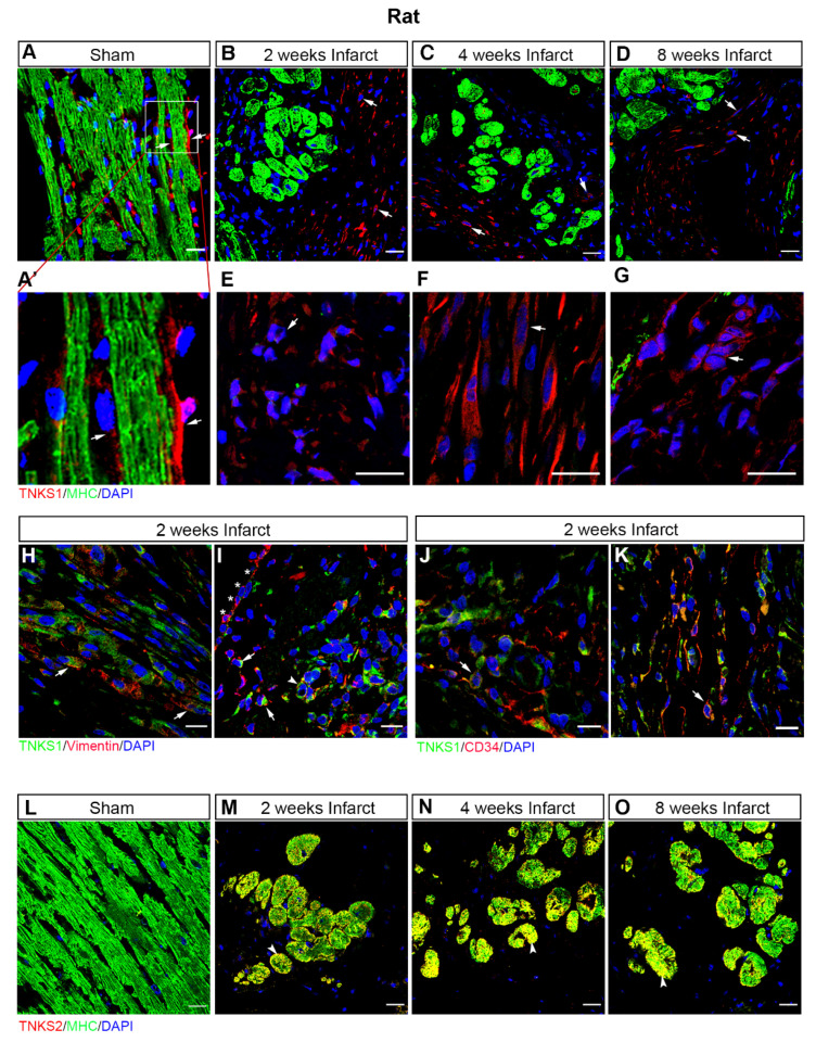
Fig. 4
Ischemic injury stimulates TNKS1 in non-cardiomyocytes and TNKS2 in cardiomyocytes in the infarct area in rat hearts. (A–D) Representative immunohistochemical staining of TNKS1 (in red) and MHC (in green) in ventricular sections from sham-operated and MI rat hearts at 2, 4, and 8 weeks post-MI. The TNKS1-positive signal is visualized in the vasculature (arrow) in sham-operated hearts (A,A’) and in non-cardiomyocytes (examples indicated by arrows) in the infarct area at the examined time points (B–D). (A’) Inset: magnified image of TNKS1-positive cells within the vasculature. (E–G) Representative images of immunohistochemical staining showing the TNKS1-positive signal in granulocytes-like cells (E), fibroblasts (F), and endothelium-like cells (G) indicated by the arrow in the infarct area. (H,I) Representative immunohistochemical staining of TNKS1 (in green) and vimentin (in red) showing accumulation of TNKS1 in vimentin-positive fibroblasts (arrow in H), EMT cells (arrow in I), and vascular endothelial cells (arrowhead in I) in the infarct area at 2 weeks post-MI. Stars (* in I) indicate the epicardium. (J,K) Representative immunohistochemical staining of TNKS1 (in green) and CD34 (in red) showing accumulation of TNKS1 in CD34-positive (arrow) vascular endothelial cells (J) and circulating cells (K) in the infarct area at 2 weeks post-MI. (L–O) Representative immunohistochemical staining of TNKS2 (in red) and MHC (in green) in ventricular sections from sham-operated and infarcted rat hearts at 2, 4, and 8 weeks post-MI. The co-localization of TNKS2 and MHC is visualized in cardiomyocytes (arrowhead) in the infarct area at the examined time points. Scale bars: 20 μm.