
|
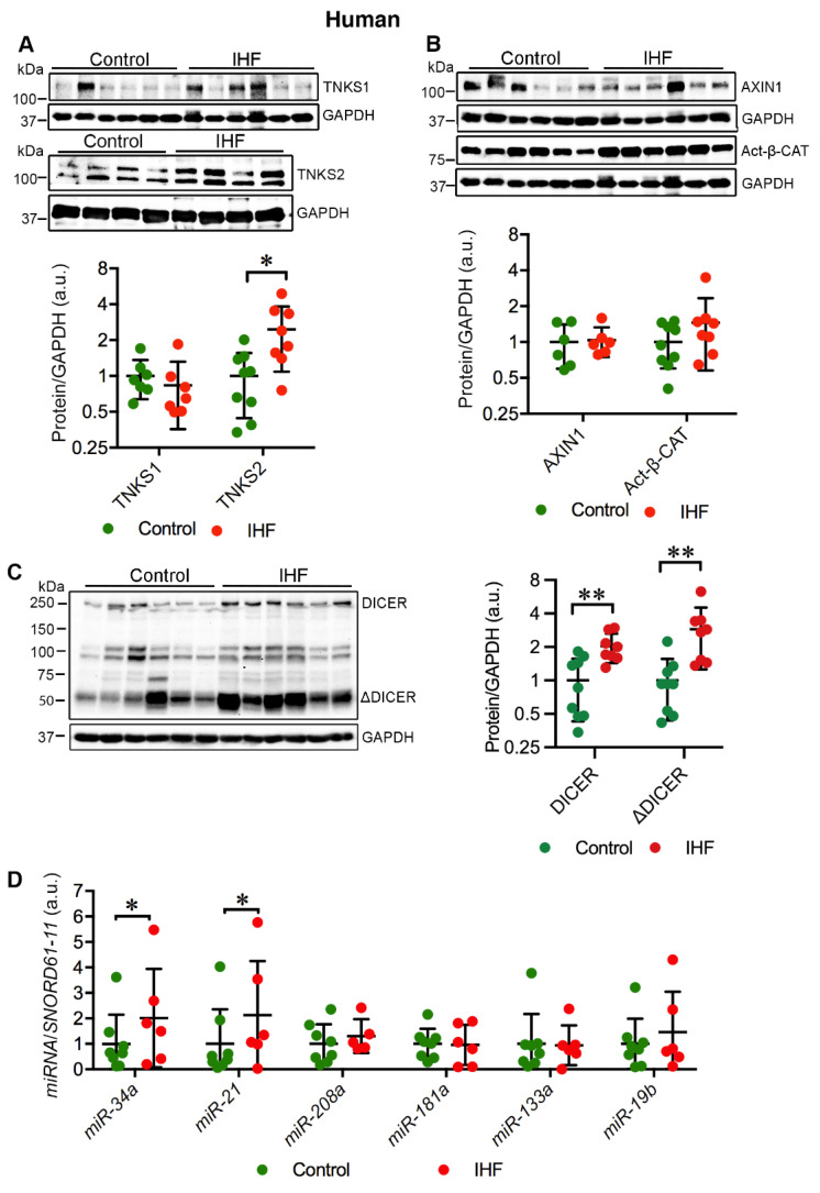
Fig. 1
Cardiac TNKS2 and DICER are stabilized in ischemic heart failure (IHF) patients. (A) Representative immunoblots of TNKS1 and TNKS2 in the left ventricle (LV) of IHF and healthy controls. (B) Representative immunoblots of AXIN1 and active β-catenin (Act-β-CAT) in the LV of IHF and healthy controls. (C) Representative immunoblots of DICER and truncated DICER (ΔDICER) in the LV of IHF and healthy controls. The graphs represent quantifications of three replicate blots for the indicated proteins relative to GAPDH. The results are presented as fold change compared to controls. The blurry or oversaturated bands were excluded from the quantification. Controls: TNKS1, n = 7; TNKS2, n = 9; AXIN1, n = 6; Act-β-CAT, n = 9; DICER, n = 9; ΔDICER, n = 9; IHF: TNKS1, n = 7; TNKS2, n = 8; AXIN1, n = 6; Act-β-CAT, n = 8; DICER, n = 8; ΔDICER, n = 8. (D) qRT-PCR analysis of miRNA expression in the LV of IHF patients (n = 6) and healthy controls (n = 8). Data are presented as mean ± SD. Two-sample t-test: * p < 0.05, ** p < 0.01.