
|
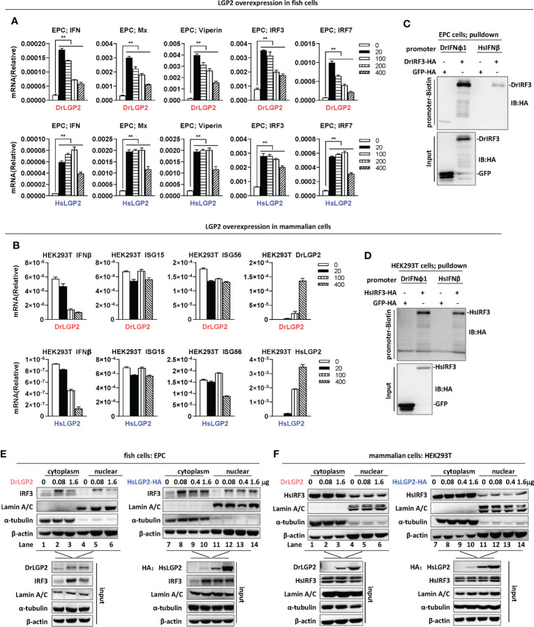
Fig. 2
Overexpression of zebrafish or human LGP2 induces IFN response in fish cells but not in mammalian cells (A, B) RT-PCR analysis of transcriptional levels of IFN and ISGs induced by DrLGP2 and HsLGP2 in EPC cells (A) and HEK293T cells (B). EPC cells (A) and HEK293T cells (B) seeded overnight in 12-wells plates were transfected with DrLGP2 or HsLGP2 at increasing doses (0, 20, 10, 200, 400 ng) for 24 h, followed by RT-PCR detection of cellular gene transcription. P values were calculated using ANOVA. **P < 0.01. (C, D) DNA pull-down assays verified the binding of DrIFNφ1 and HsIFNβ promoter DNA to DrIRF3 in EPC cells (C) and HsIRF3 in HEK293T cells (D). EPC cells (C) and HEK293T cells (D) seeded in 10 cm dishes were transfected with DrIRF3-HA (C) or HsIRF3-HA (D). GFP-HA was transfected in parallel as control. 24 h later, cells were lysed. One-tenth of cell lysates were taken as input, and the remaining was incubated overnight with 100 ng biotinylated DrIFNφ1 promoter DNA (-596 to +38) (C) or HsIFNβ promoter DNA (-338 to +93) (D). The DNA-bound protein complexes were detected by western blots with anti-HA antibody. (E, F) overexpression of LGP2 increased nuclear IRF3 protein levels in fish cells but not in mammalian cells. EPC cells (E) and HEK293T cells (F) seeded in 5 cm dishes were transfected with DrLGP2 or HsLGP2 at increasing doses. 24 h later, cells were collected for nuclear and cytoplasmic separation, followed by western blot analyses of the indicated proteins using corresponding antibodies. The expression of Lamin A/C and α-tubulin verified the successful separation of nuclear and cytoplasmic lysates.