Fig. 6
Image
Figure Caption
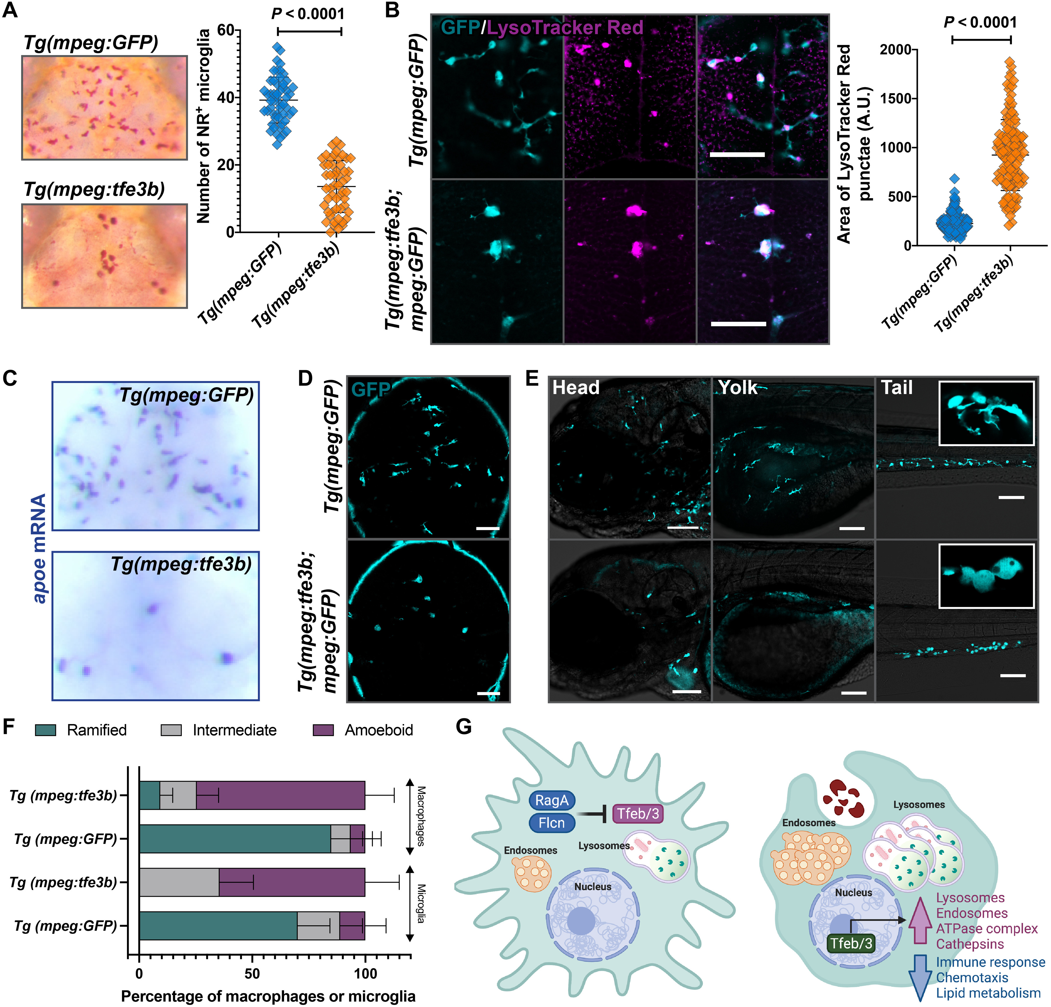
Fig. 6. Overexpression of tfe3b in the macrophage lineage disrupts microglia number and morphology as in rraga mutants.
Comparison of macrophages and microglia between animals overexpressing Tfe3b in the macrophage lineage, Tg(mpeg:tfe3b), and controls, Tg(mpeg:GFP), using (A) neutral red assay and quantification and (B) LysoTracker Red assay and quantification. Graphs show mean + SD; significance was determined using parametric unpaired t test. (C) apoe in situ hybridization and (D and E) live imaging with the mpeg:GFP transgene in (D) the brain and (E) the head, yolk, and tail regions of Tg(mpeg:tfe3b) and Tg(mpeg:GFP) larvae. Insets show magnified views of cell morphology. (F) Quantification of amoeboid morphology of microglia and macrophages. Graph shows mean + SD; significance was determined using nonparametric Mann-Whitney U test. Scale bars, 50 μm. The number of animals analyzed for each experiment is listed in table S1; all the panels are representative of at least two independent experiments. (G) Schematic summarizing the lysosomal regulatory circuit in microglia and macrophages.
Figure Data
Acknowledgments
This image is the copyrighted work of the attributed author or publisher, and
ZFIN has permission only to display this image to its users.
Additional permissions should be obtained from the applicable author or publisher of the image.
Full text @ Sci Adv

