Fig. 5
Image
Figure Caption
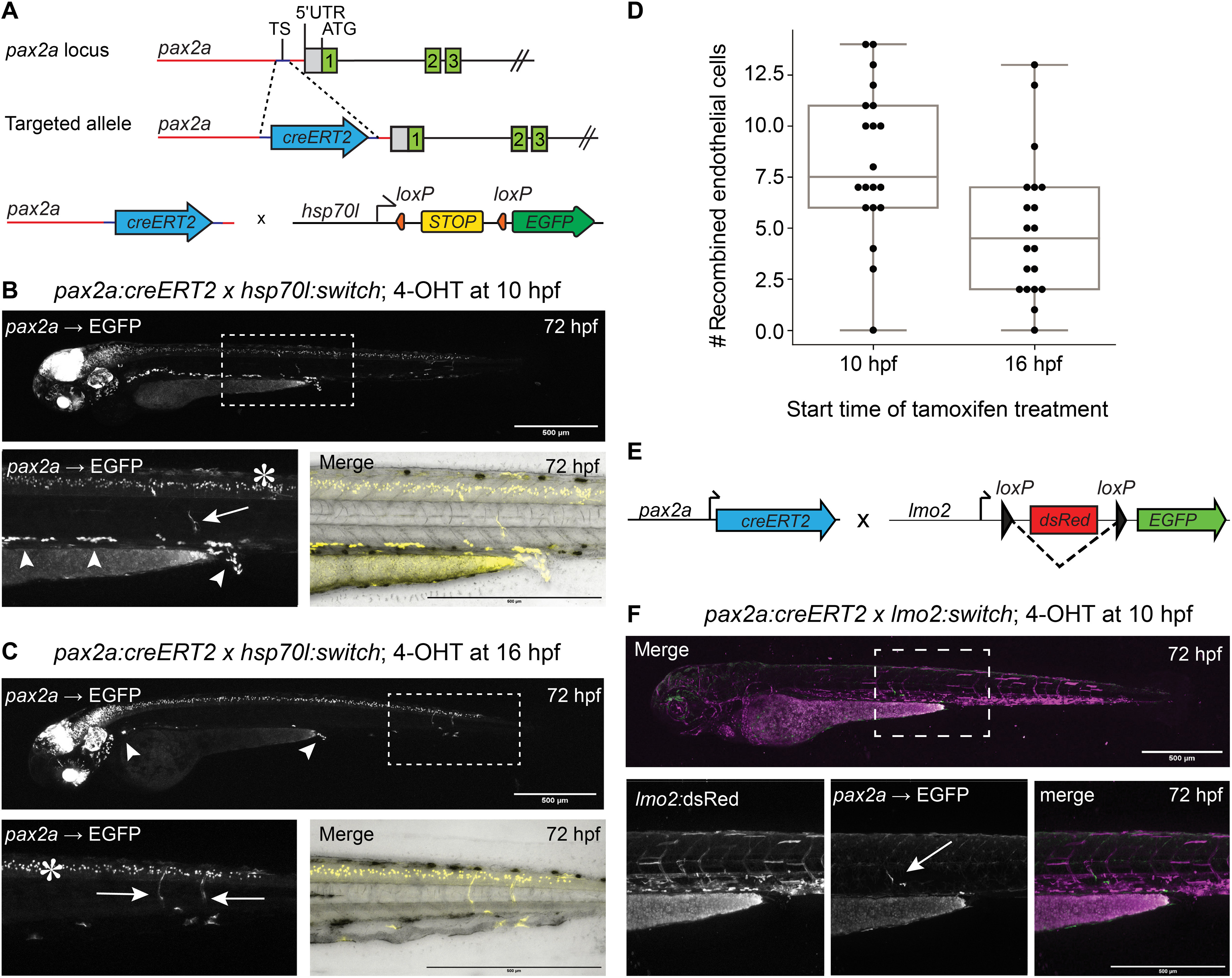
Fig. 5. Lineage tracing of pax2a-expressing cells labels endothelial cells.
(A) Schematic representation of the pax2a:cre:ERT2 knock-in line. pax2a:creERT2 zebrafish were crossed with hsp70l:Switch zebrafish, and the resulting embryos were treated with 4-OHT from 10 to 24 hpf or 16 to 36 hpf and heat-shocked at 72 hpf for 1 hour. TS, transcriptional start. Several pax2a:creERT2 switched EGFP-positive endothelial cells (arrows) can be observed in zebrafish treated with 4-OHT from 10 (B) or 16 (C) hpf. Arrowheads point to pronephron cells, and asterisks indicate spinal cord neurons. Recombined circulating blood cells can be seen in movie S2. 5′UTR, 5′ untranslated region. (D) Quantification shows a decrease in the number of recombined endothelial cells in the animals treated starting at 16 hpf compared with those treated starting at 10 hpf. (E) Schematic representation of the pax2a:creERT2-mediated recombination of an lmo2:Switch line. (F) Recombined endothelial cells (arrow) are observed in the trunk and tail of lmo2:Switch larvae. Scale bars, 500 μm.
Figure Data
Acknowledgments
This image is the copyrighted work of the attributed author or publisher, and
ZFIN has permission only to display this image to its users.
Additional permissions should be obtained from the applicable author or publisher of the image.
Full text @ Sci Adv

