
Image
|
Figure Caption
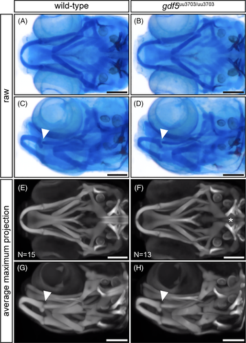
Fig. 6
Optical projection tomography reveals no craniofacial skeletal defects in gdf5uu3703/uu3703 zebrafish at 9 dpf. (A-D) Examples of raw images of single 9 dpf zebrafish heads captured during OPT. (E-H) Maximum projection views of averaged 3D models generated by OPT analysis of 15 wild-type and 13 gdf5 mutant zebrafish at 9 dpf. Arrowheads indicate the jaw joint between Meckel's cartilage and the palatoquadrate. The asterisk highlights the reduced anterior notochord ossification in gdf5uu3703/uu3703 fish relative to wild-types. Scale bars: 150 μm
Figure Data
Acknowledgments
This image is the copyrighted work of the attributed author or publisher, and
ZFIN has permission only to display this image to its users.
Additional permissions should be obtained from the applicable author or publisher of the image.
Full text @ Dev. Dyn.