IMAGE

Fig. 6

ID
ZDB-IMAGE-220824-10
Source
Figures for Karampelias et al., 2022
Image
Figure Caption

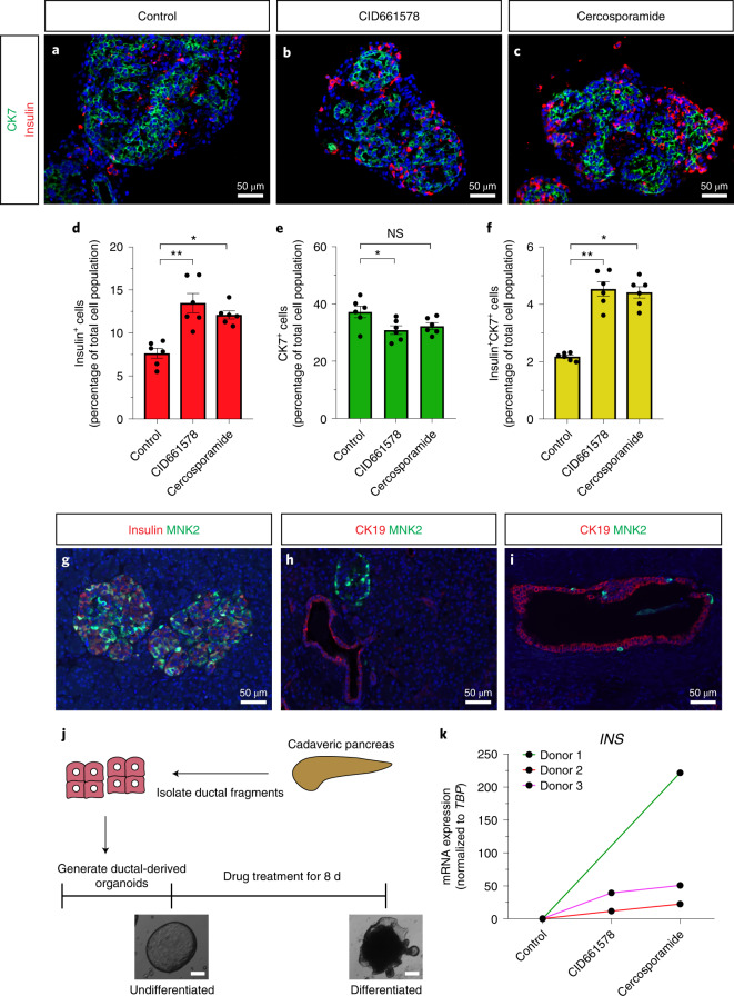
Fig. 6 CID661578/cercosporamide treatment increases β-cell differentiation in ductal cells from neonatal pigs and human organoids.

af, Images of neonatal pig islets treated with DMSO (a), CID661578 (b) or cercosporamide (c) and stained for insulin (red) and the ductal cell marker CK7 (green). Quantification results showed that treatment with either CID661578 or cercosporamide increased the number of insulin+ β-cells (d), decreased the number of CK7+ duct cells (e) and increased the number of double-positive (insulin+CK7+) cells (f); n = 6; **P = 0.0041 and *P = 0.0116 (d); *P = 0.0401 and P = 0.1671 (NS, not significant) (e); **P = 0.0034 and *P = 0.0137 (f). A Kruskal–Wallis test followed by Dunn’s multiple comparisons test was used to assess significance for df. Data are presented as mean values ± s.e.m. gi, Images of human pancreatic sections from different donors stained for MNK2, with insulin used as a marker of β-cells and CK19 used to mark the pancreatic duct. Similar results have been reproduced in stainings from pancreatic sections of multiple human donors. j,k, Schema showing the procedure for generating and treating human ductal-derived organoids (j). Brightfield images of representative examples of human ductal-derived organoids before differentiation and after treatment with cercosporamide are shown; scale bar, 200 µm. INS mRNA expression is shown in k for three different organoid preparations (that is, from three different donors) for cercosporamide and two for CID661578. The experiment was reproducible in at least two different organoid preparations.

Source data

Acknowledgments
This image is the copyrighted work of the attributed author or publisher, and ZFIN has permission only to display this image to its users. Additional permissions should be obtained from the applicable author or publisher of the image. Full text @ Nat. Chem. Biol.