
Image
|
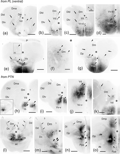
Figure Caption
Fig. 17
(a–o) Photomicrographs of transverse sections through the telencephalic lobes showing labeled cells (black arrowheads) and fibers (arrows) after application of DiI to the ventral part of the hypothalamic posterior lobe (a–g) and to the posterior tuberal nucleus (PTN) (h–o). Open arrowheads point to the sulcus ypsiloniformis. Numbers 1–3 indicate Dm subdivisions. Asterisk, midline ventricle. For abbreviations, see the list. All photomicrographs are negative images of fluorescent data. Scale bars: 200 μm (a–c, e, g, h–i, l, n); 100 μm (j–k); 50 μm (d, f)
Acknowledgments
This image is the copyrighted work of the attributed author or publisher, and
ZFIN has permission only to display this image to its users.
Additional permissions should be obtained from the applicable author or publisher of the image.
Full text @ J. Comp. Neurol.