
Image
|
Figure Caption
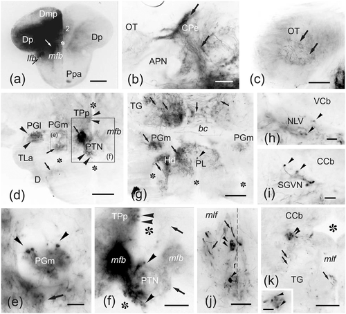
Fig. 15
(a–k) Photomicrographs of transverse sections of the zebrafish brain ordered from rostral to caudal showing cells (arrowheads) and fibers (arrows) labeled after application of DiI to Dc in a brain en-block sectioned at commissural level. (a) Panoramic view of a section of the telencephalic lobes caudal to the application point. Note that Dp and Dm2 were heavily labeled, probably en-passant, through the various fibers bundles traversing Dc between major telencephalic tracts and more superficial pallial areas. Ipsilateral is to the left. (b) Section through the ipsilateral pretectum showing dense innervation of the central pretectal nucleus (CPe) by labeled fibers. (c) Section through the rostral optic tectum showing labeled varicose fibers. (d–f) Section through the hypothalamus-posterior tuberal area (d) and details (e–f) showing labeled cells and fibers in the ipsilateral PGl, PGm, and PTN. (g) Sections through the caudal hypothalamus and midbrain tegmentum showing bilateral labeled projections to the PGm, the hypothalamic posterior lobe and the tegmentum, and ipsilateral projection to Hv. (h–j) Details showing labeled cells in the lateral valvular nucleus (h), the secondary gustatory/visceral nucleus (i), and the superior raphe nucleus (j). (k) Section at the level of the trigeminal nerve exit showing a group of labeled cells in the ipsilateral dorsal rhombencephalic tegmentum and fibers in the reticular area. Asterisk, ventricle. For abbreviations, see the list. All photomicrographs are negative images of fluorescent data. Scale bars: 200 μm (a, c–d, g, k); 100 μm (b, f); 50 μm (e, h–j, inset in k)
Acknowledgments
This image is the copyrighted work of the attributed author or publisher, and
ZFIN has permission only to display this image to its users.
Additional permissions should be obtained from the applicable author or publisher of the image.
Full text @ J. Comp. Neurol.