
Image
|
Figure Caption
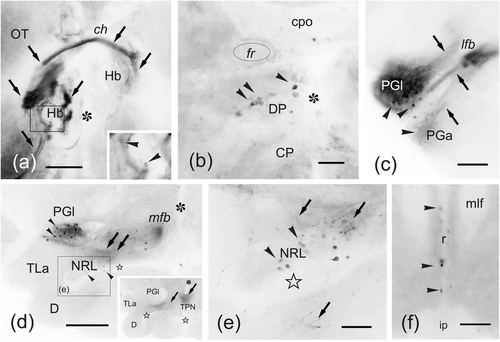
Fig. 10
(a–f) Photomicrographs of transverse sections through the zebrafish brain showing labeling in extratelencephalic regions after application of DiI to precommissural Dl. Arrowheads: cell bodies. Arrows: fibers and tracts. (a) Transverse section through the epithalamus showing occasional labeled cells (inset) and fibers crossing through the habenular commissure to reach the contralateral habenula. (b) Section showing labeled cells in the dorsal posterior nucleus of the thalamus. (c–e) Photomicrographs and details of sections of the hypothalamus-posterior tubercle, showing labeled cells and fibers from the ipsilateral Dl in the lateral preglomerular nucleus (c–d) and nucleus of the lateral recess (e). (f) Section of the isthmus showing labeled cells in the superior raphe dorsally to the interpeduncular nucleus. Arrowheads point to labeled cells, arrows to fibers. Asterisk: third ventricle (a, b) and hypothalamic lateral recess (d–e). Outlined stars: inferior and posterior lobe recesses. For abbreviations, see the list. All photomicrographs are negative images of fluorescent data. Scale bars: 200 μm (d); 100 μm (a, c, f); 50 μm (b, e)
Acknowledgments
This image is the copyrighted work of the attributed author or publisher, and
ZFIN has permission only to display this image to its users.
Additional permissions should be obtained from the applicable author or publisher of the image.
Full text @ J. Comp. Neurol.