
Image
|
Figure Caption
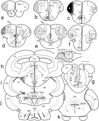
Fig. 8
(a–k) Schematic drawings of transverse sections of the zebrafish brain showing the distribution of cells and fibers labeled after application of DiI to the lateral zone of the pallium (Dl). The level of sections from rostral to caudal is indicated in a lateral view of the brain. Black circles, retrogradely labeled cells. Small dots and lines, labeled fibers and tracts. Shaded areas in (a–e) represent the areas with dense labeling by the tracer from the point of application of the small DiI crystal (white star in c). Ipsilateral is at the left. Scale bar for sections: 500 μm
Acknowledgments
This image is the copyrighted work of the attributed author or publisher, and
ZFIN has permission only to display this image to its users.
Additional permissions should be obtained from the applicable author or publisher of the image.
Full text @ J. Comp. Neurol.