
Image
|
Figure Caption
Fig. 5
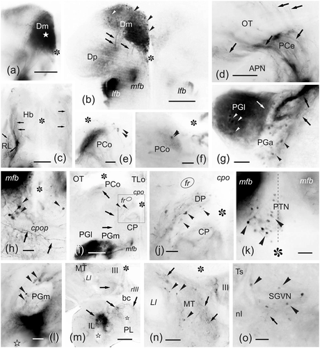
(a–o) Photomicrographs of transverse sections of the zebrafish brain showing labeled structures after application of DiI to the medial zone of the pallium (Dm1-2). The correspondence with the levels of Figure 4 is indicated. (a) Section close to the application point (white star) (see Figure 4b). (b) General view of the posterior telencephalic lobe showing numerous labeled cells in caudal Dm (see Figure 4d). (c) Detail of the habenular region showing labeled varicose fibers in the ipsilateral rostrolateral nucleus and in a lateral plexus in both habenulae (see Figure 4e). (d) Labeled varicose fibers coursing through the ipsilateral central pretectal nucleus (see Figure 4d) and entering the optic tectum. (e–f) Detail of labeled cell bodies and fibers in the ipsi- (e) and contralateral (f) paracommissural pretectal nucleus (see Figure 4g). (g) Section through the anterior and lateral preglomerular nuclei, showing numerous labeled cells and fibers (see Figure 4g). (h) Detail of a small group of retrogradely labeled cell bodies close to the medial forebrain bundle and dorsal to the postoptic commissure. (i–j) General view (i) and detail (j) of the ipsilateral dorsal posterior thalamic nucleus showing a few cell bodies and fibers labeled. Note also the dense neuronal labeling in the medial and lateral preglomerular nuclei. (k) Detail showing retrogradely labeled cell bodies in the ipsilateral side of the posterior tuberal nucleus (the broken line indicates the midline) (see Figure 4h). (l) Detail showing a few cell bodies and fibers labeled in the medial preglomerular nucleus (see Figure 4h). (m) Panoramic view showing numerous labeled fibers in the ventral mesencephalic tegmentum around the third nerve root, and in the hypothalamic inferior and posterior lobes (see Figure 4i). (n) Detail of labeled cell bodies and fibers between the lateral lemniscus and the oculomotor nucleus in the medial mesencephalic tegmentum. (o) Retrogradely labeled cell bodies in the rostral part of the secondary gustatory-visceral nucleus (see Figure 4j). Arrowheads point to cell bodies. Arrows point to fibers and fiber tracts. For abbreviations, see the list. Asterisk: ventricle. Outlined star: recesses of the inferior lobes. Medial is to the right. All photomicrographs are negative images of fluorescent data. Scale bars: 200 μm (a–b, i, m); 100 μm (c–d, n); 50 μm (e–h, j–l, o)
Acknowledgments
This image is the copyrighted work of the attributed author or publisher, and
ZFIN has permission only to display this image to its users.
Additional permissions should be obtained from the applicable author or publisher of the image.
Full text @ J. Comp. Neurol.