
Image
|
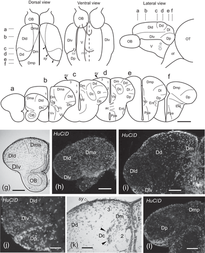
Figure Caption
Fig. 1
Upper panel, schematic drawings of dorsal, ventral, and lateral views of the adult zebrafish telencephalon showing the external distribution of the main pallial areas considered in this study and the location of internal tracts observed thanks to certain transparency of the tissue (gray lines). Middle panel (a–f), schemes of transverse sections of the telencephalon showing the location of the different pallial and subpallial regions considered in this study. The levels of sections are indicated with dashes in the upper panel. Lower panel (g–l), selected photomicrographs of Nissl staining and HuC/D immunocytochemistry of transverse sections of the zebrafish telencephalon at rostral (g, h) precommissural (i, j), commissural (k), and postcommissural (k) levels showing cytoarchitectonical features of the telencephalic areas used in this study. For abbreviations, see the list. Scale bars: 200 μm (a–f, h–i, l) and 100 μm (g, j–k)
Acknowledgments
This image is the copyrighted work of the attributed author or publisher, and
ZFIN has permission only to display this image to its users.
Additional permissions should be obtained from the applicable author or publisher of the image.
Full text @ J. Comp. Neurol.