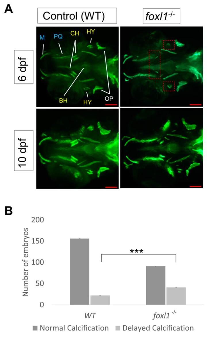
IMAGE

Figure 2

ID
ZDB-IMAGE-220801-137
Source
Figures for Hawkey-Noble et al., 2022
Image
Figure Caption

Figure 2

Calcein staining illustrating the impact of foxl1 loss on craniofacial development and calcification. (A) Embryos of WT aged 6 and 10 dpf (n = 173 and 99, respectively) as well as those of foxl1-/- (n = 116 and 65, respectively). WT embryos at both 6 and 10 dpf exhibit normal craniofacial development and calcification of all jaw structures. foxl1-/- embryos show a delayed calcification in the ceratohyal and hyomandibula (red boxes) at 6 dpf yet appear mostly recovered by 10 dpf. (B) The proportion of embryos with delayed calcification in foxl1 mutants is statistically significant (Fisher’s *** p = 0.0001) when compared to wildtype siblings. PQ, palatoquadrate; M, Meckel; HY, hyomandibula (hyosympletic); BH, basihyal; OP, opercula; CH, ceratohyal. Scale bars are 100 µm.

Figure Data
Acknowledgments
This image is the copyrighted work of the attributed author or publisher, and ZFIN has permission only to display this image to its users. Additional permissions should be obtained from the applicable author or publisher of the image. Full text @ Genes (Basel)