
|
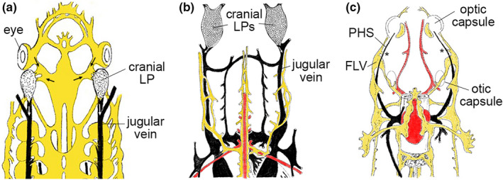
FIGURE 5 Examples of lymphatic anatomy in cartilaginous and bony fish. Traced drawings depicting the anterior lymphovenous networks in (a) jawless fish (lamprey, after metamorphosis), (b) cartilaginous fish (shark) and (c) bony fish (trout). Dorsal views, lymphatic vessels depicted in yellow, veins in black, and arteries in red. (a, b) Cranial lymph propulsor (LP) and jugular veins are noted. Arrows indicate direction of flow. (c) Primary head sinus (PHS) and facial lymphatic vessel (FLV) assigned based on similar anatomical positions in larval zebrafish. Traced sketches after Kampeier, 1969 with permission