
|
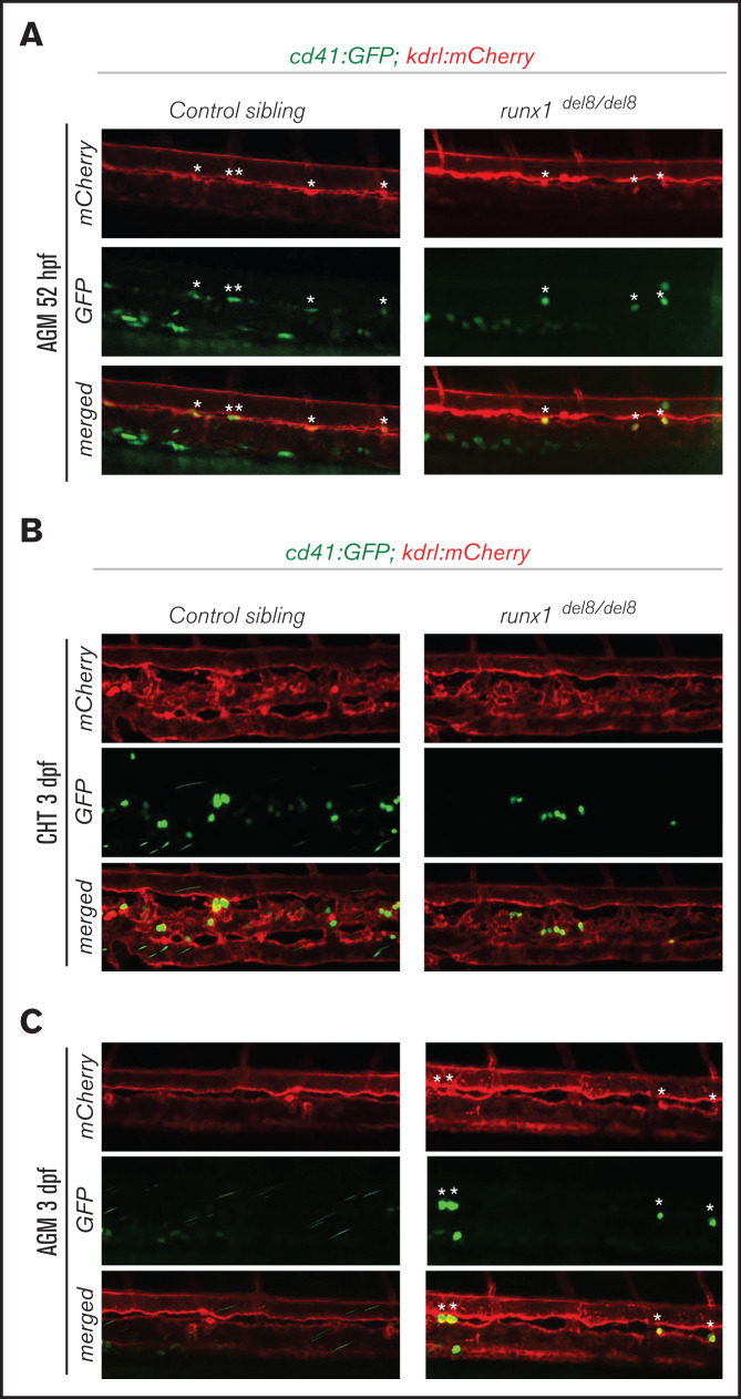
Figure 2.
Development of cd41:GFP+ hematopoietic precursors in runx1del8/del8 AGM and CHT. (A) Snapshots of the AGM region of runx1del8/del8 and control sibling Tg(cd41:GFP); Tg(kdrl:mCherry) at 52 hours postfertilization (hpf), obtained from time-lapse recordings on a confocal microscope (supplemental Movies 1 and 2). cd41:GFP+ mCherry+ double-positive cells are indicated with white asterisks. (B-C) Live confocal imaging of the CHT (B) and AGM (C) regions in the Tg(cd41:GFP); (B) Tg(kdrl:mCherry) embryos at 3 dpf. cd41:GFP+ cells are found in the CHT. (C) Residual cd41:GFP+ cells (white asterisks) are still detectable in the AGM of runx1del8/del8.