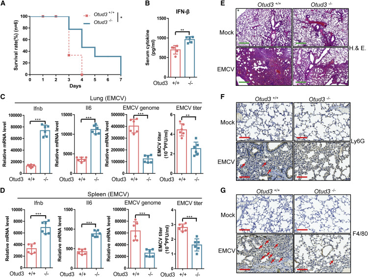
Fig. 3 Figure 3. OTUD3-deficient mice are more resistant to EMCV infection (A) Survival (Kaplan-Meier curve) of Otud3+/+ and Otud−/− mice (n = 6 per group) at various times after intraperitoneal (i.p.) injection with EMCV (1 × 10⁶ plaque-forming units [PFU] per mouse). (B) ELISA of IFN-β in serum from Otud3+/+ and Otud3−/− mice (n = 5 per group) given i.p. injection of EMCV (1 × 10⁶ PFU per mouse) for 12 h. (C and D) qRT-PCR analysis of Ifnb, IL6, EMCV mRNA, and plaque assays for EMCV titers in the lungs (C) and spleens (D) from Otud3+/+ and Otud3−/− mice given i.p. injection of EMCV (1 × 10⁶ PFU per mouse) for 24 h. (E–G) Microscopy of H&E-stained (E), Ly6G-stained (F), or F480-stained lung sections from Otud3+/+ and Otud3−/− mice treated with PBS (mock) or EMCV (1 × 10⁶) for 48 h. Red arrows indicate positive cells. E Scale bar, 500 μm; (F and G) scale bar, 100 μm. ∗p < 0.05, ∗∗p < 0.01, and ∗∗∗p < 0.001, using unpaired Student’s t test (B–D) or log rank (Mantel-Cox) test (A). Data based on one representative experiment performed in three biological replicates from at least three independent experiments (mean ± SD) or representative data (A, E, F, and G).
Image
Figure Caption
Acknowledgments
This image is the copyrighted work of the attributed author or publisher, and
ZFIN has permission only to display this image to its users.
Additional permissions should be obtained from the applicable author or publisher of the image.
Full text @ Cell Rep.

