
|
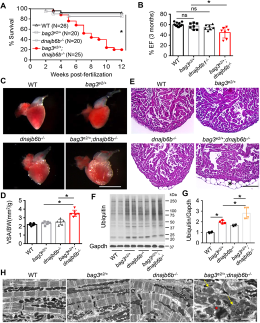
Fig. 2
Genetic interaction between bag3e2/+ and dnajb6b−/− in adult zebrafish heart. (A) Survival (in %) of bag3e/+;GB411−/− double-mutant fish compared to single-mutant and WT control fish; n=25-40, log-rank test. (B) Ejection fraction (EF) (in %) of bag3e2/+;dnajb6b−/− double mutants compared to single mutants and WT controls at 3 months post fertilization; n=6-8, one-way ANOVA. (C-E) Bright-field images of dissected hearts (C) show significantly enlarged ventricular surface area (VSA) in bag3e2/+;dnajb6b−/− double-mutant fish. VSA normalized to body weight (BW) index is plotted in D. H&E staining of heart ventricles (E) highlights the obvious lipid accumulation phenotype (asterisk); n=3-4, one-way ANOVA. Scale bars: 1 mm (C), 100 µM (E). (F,G) Western blot (F) and quantification analysis (G) of ubiquitylated protein levels in heart lysates of bag3e2/+;dnajb6b−/− double-mutant fish compared to those of single-mutant and WT control fish at 3 months; n=4, one-way ANOVA. (H) Confirmative TEM images, showing mitochondrial swelling (arrows) and myofibril degeneration phenotypes (asterisk) the bag3e2/+;dnajb6b−/− double-mutant fish hearts at 3 months. Scale bar: 2 µM. *P<0.05; ns, not significant.