
IMAGE
Fig. 5
- ID
- ZDB-IMAGE-220622-72
- Publication
- Nguyen et al., 2022 - Dynamics of the Zebrafish Skeleton in Three Dimensions During Juvenile and Adult Development
- All Figures
- Figures for Nguyen et al., 2022
Image
|
Figure Caption
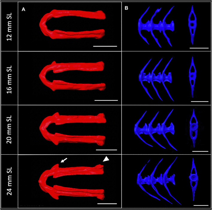
Fig. 5
Shape change of the lower jaw and caudal vertebrae. (A), Segmented lower jaws from 12, 16, 20, and 24 mm SL individuals, viewed from the ventral perspective. In the largest individuals, note the pronounced anguloarticular prominence (arrow) and posterior end of lower jaw (arrowhead). (B), Segmented first three caudal vertebrae from 12, 16, 20 and 24 mm SL individuals, viewed from a lateral perspective. Scale bars, 0.5 mm.
Acknowledgments
This image is the copyrighted work of the attributed author or publisher, and
ZFIN has permission only to display this image to its users.
Additional permissions should be obtained from the applicable author or publisher of the image.
Full text @ Front. Physiol.