
|
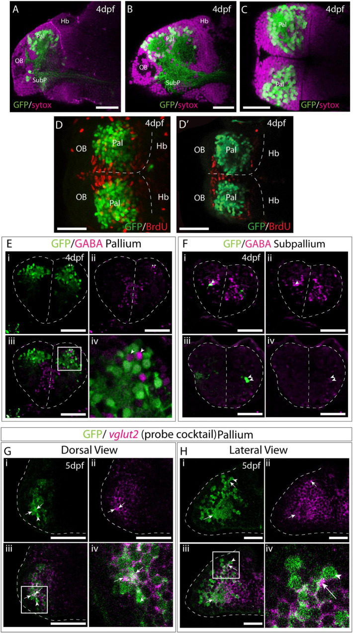
Fig. 7
Et(gata2:EGFP)bi105 is expressed in post-mitotic glutamatergic pallial neurons. Sagittal (A,B) and horizontal (C) sections through a 4dpf Et(gata2:EGFP)bi105 brain stained with sytox (magenta) to label cell nuclei and anti-EGFP (green). EGFP expression is largely excluded from the dorsal and medial ventricular zones of the telencephalon. (D,D′) Dorsal views of a 4dpf Et(gata2:EGFP) bi105 brain labeled with anti-BrdU (red) and anti-GFP (green) antibodies. (D) z-projection and (D′) shows a single coronal z-slice through the same brain. BrdU is excluded from the EGFP+ neurons in the dorsal telencephalon indicating that these cells are likely to be non-proliferative and postmitotic. (E–F) Transverse sections through the telencephalon of a 4dpf Et(gata2:EGFP) bi105 brain stained with anti-GABA (magenta) to label GABAergic cells and anti-GFP (green). EGFP+ cells in the pallium (E) are for the most part GABA-. Gaps where GABAergic pallial interneurons intermingle with EGFP+ pallial neurons are visible [arrowheads in panel (Ei)]. In subpallial telencephalic domains (F) some cells do co-express EGFP and GABA [arrowheads in panels (Fi–iv)]. Panels (Fi–iv) are different z-sections through the same Et(gata2:EGFP) bi105 brain showing subpallial EGFP+ neurons on both sides of the brain. Dorsal (G) and lateral (H) views of Et(gata2:EGFP) bi105 brains at 5dpf labeled with FISH using a “cocktail” probe for vglut2a/slc17a6b and vglut2b/slc17a6a (magenta) and antibody labeling with anti-GFP (green) show overlap of vglut2a and EGFP within the telencephalic neuropil (arrows) associated with EGFP+ cells (arrowheads). (Giv) High-magnification view of dorsal telencephalic neurons in boxed region in panel (Giii). (Hiv) High-magnification view of dorsal telencephalic neurons in boxed region in panel (Hiii). Scale bars: 50 μm.