
Image
|
Figure Caption
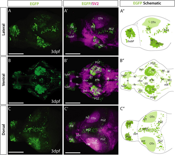
Fig. 2
Et(gata2:EGFP)bi105 line EGFP expression at 3 dpf. Lateral (A,A′), ventral (B,B′) and dorsal (C,C′) views of a 4 dpf Et(gata2:EGFP)bi105 larvae labeled with anti-EGFP (green) and anti-SV2 antibodies (magenta). Major areas of EGFP expression are annotated in the schematics on the right (A″,B″,C″), these include: pallium, subpallial nucleus, pretectum, hypothalamic areas, optic tectum and hindbrain areas, among others. Scale bars: 100 μm.
Acknowledgments
This image is the copyrighted work of the attributed author or publisher, and
ZFIN has permission only to display this image to its users.
Additional permissions should be obtained from the applicable author or publisher of the image.
Full text @ Front. Neuroanat.