
|
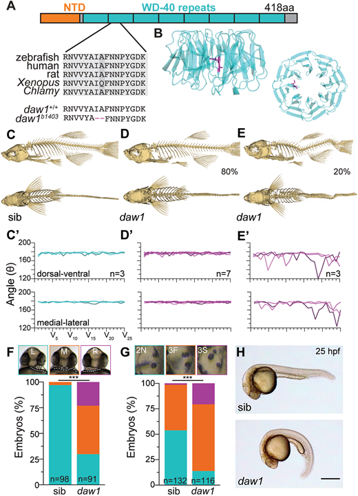
Fig. 1
daw1b1403 mutants exhibit motile cilia-associated defects. (A) The 418 amino acid Daw1 protein with short N-terminal domain (NTD) and large β-propeller composed of WD40 repeats. daw1b1403 deletes two conserved residues, I140 and A141. (B) Human DAW1 X-ray structure (Protein Data Bank: 5NNZ) showing I140 and A141 location. (C-E) 3D reconstitutions of µCT data. Most daw1b1403 mutants showed normal spines (D), but 20% exhibited curves (E), compared with control (C). (C′-E′) Quantitation of curvature showed curved daw1b1403 mutants had a non-stereotyped pattern (E′), compared with control (C′) and non-curved daw1b1403 mutants. (F) Top: representative image of 25 hpf larvae showing the heart (dotted oval) on top of the yolk (dotted line). Bottom: daw1b1403 mutants exhibited randomized heart laterality including left (L; green), middle (M; orange) and right (R; magenta)-facing hearts (n=91 mutants and 98 sib controls; ***P=4.84×10⁻²¹, chi square). (G) Top: otic vesicles at 25 hpf showed abnormal phenotypes in daw1b1403 mutants [2N (green) – two normal; 3F (orange) – three otoliths, posterior two fused; 3S (magenta) – three separate otoliths. Bottom: quantification of n=116 mutants and 132 sibs; ***P=1.31×10⁻¹², chi square. (H) CTD in daw1b1403 mutants was fully penetrant at 25 hpf. Scale bar: 0.5 mm (H).