
Image
|
Figure Caption
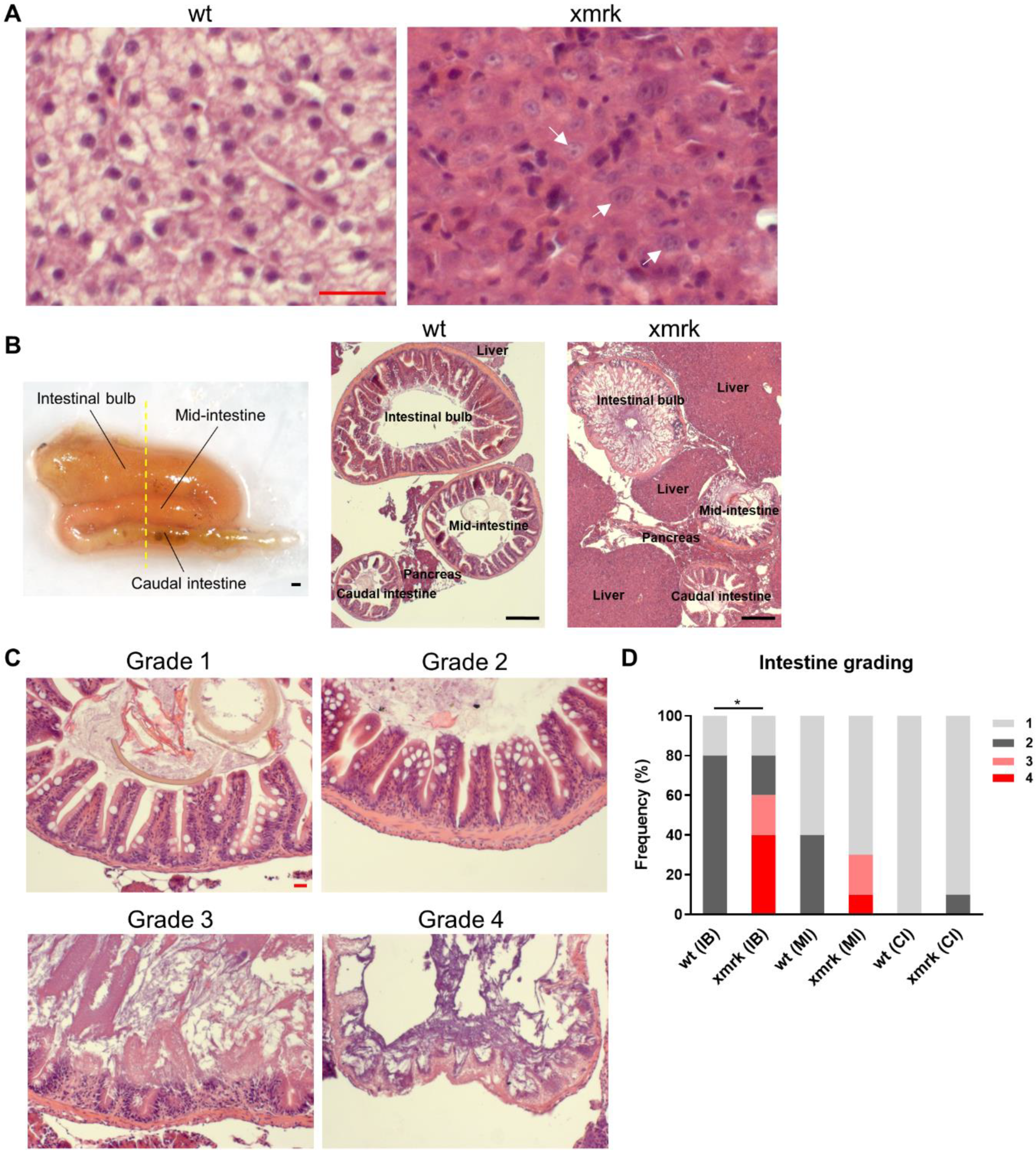
Fig. 1
Figure 1. Disruption of intestine morphology after 6 weeks of HCC induction. (A) Representative H&E images showing the normal liver in wild-type (wt) fish and the HCC liver in xmrk fish. It was found that 100% of xmrk livers progressed into HCC after 6 weeks of doxycycline treatment. White arrows indicate example tumor cells with large irregular nuclei and prominent nucleoli. (B) Dissected intestine showing the folding of intestine into three segments (left panel). Intestine samples were sectioned transversely in order to view all three segments of the intestine concurrently on the same section. The yellow dashed line showed the approximate position of section. Representative H&E images taken at 50× magnification showing all three intestine segments and the surrounding liver and pancreas on the same section for wt and xmrk fish, respectively (middle and right panels). (C) Representative H&E images of the intestine taken at 200× magnification. All three segments in each wt and xmrk intestine sample were assigned a grade based on phenotype severity, with grade 1 being the least severe and grade 4 being the most severe. (D) Quantification of intestine grading percentage in wt vs. xmrk. Grade numbers are indicated in the legend according to examples in C. Scale bar in red 20 μm, black 200 μm. IB: Intestinal bulb; MI: Mid-intestine; CI: Caudal intestine. * p < 0.05.
Acknowledgments
This image is the copyrighted work of the attributed author or publisher, and
ZFIN has permission only to display this image to its users.
Additional permissions should be obtained from the applicable author or publisher of the image.
Full text @ Cells