
Fig. 6
- ID
- ZDB-IMAGE-220512-30
- Genes
- Publication
- Capon et al., 2022 - Endocardial identity is established during early somitogenesis by Bmp signalling acting upstream of npas4l and etv2
- All Figures
- Figures for Capon et al., 2022
|
Fig. 6
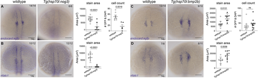
Bmp signalling is required for endocardial development. (A,B) In situ hybridisation for endocardial markers endocard:egfp (A) and nfatc1 (B) in wild-type (transgenic negative siblings) or Tg(hsp70l:nog3) embryos at the 14 somite (s) stage [heat-shock performed at tailbud stage (10 hpf)] shows significantly reduced staining of the endocardial domain upon inhibition of Bmp signalling. Quantification of the staining area of expression is shown in adjacent graphs. Quantification of GFP-positive and DAPI-positive cell number is also shown, as determined by confocal imaging of Gt(endocard:egfp), DAPI-stained embryos at 14-15 s. (C,D) Wild-type sibling controls or Tg(hsp70l:bmp2b) embryos at 14 s (heat-shocked at tailbud) show increased staining area in embryos stained for endocard:egfp (C) or nfatc1 (D). Quantification of the area of expression shown in adjacent graphs. Dorsal views are shown with anterior to the top in all images. Scale bars: 100 μm. Data are mean±s.e.m. P-values are present in graphs (unpaired two-tailed t-test). ns, not significant.