
|
Figure EV2
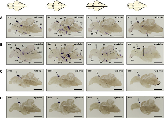
ish with ddc or asmt probe on opn4 dko mature brains show wild‐type expression patterns
A, B Same ddc expression pattern was observed in wild‐type (A) and opn4 dko (B) brains (pineal lost from the brain slices in (B) centre panels).
C, D asmt expression in wild‐type (C) and opn4 dko (D) adult brains show that asmt is not ectopically expressed. Thus, the higher asmt transcript level measured with qPCR in the opn4 dko anterior brain can be attributed to the pineal.
Data information: The horizontal line through a representation of the brain, above the section, indicates the location of the section within the brain. Abbreviations: epiphysis cerebri (pineal) [E], caudal zone of periventricular hypothalamus [Hc], inferior raphe [IR], periventricular pretectum [Pr], preoptic area [PO], posterior tuberculum [PT], paraventricular organ [PVO], suprachiasmatic nucleus [SCN], superior raphe [SR]. Note that the duration of the colorimetric development of the ishs in this figure is not exactly the same. Scale bar: 1.0 mm.