IMAGE

Figure 9.

ID
ZDB-IMAGE-220505-63
Source
Figures for Egashira et al., 2022
Image
Figure Caption

Figure 9.

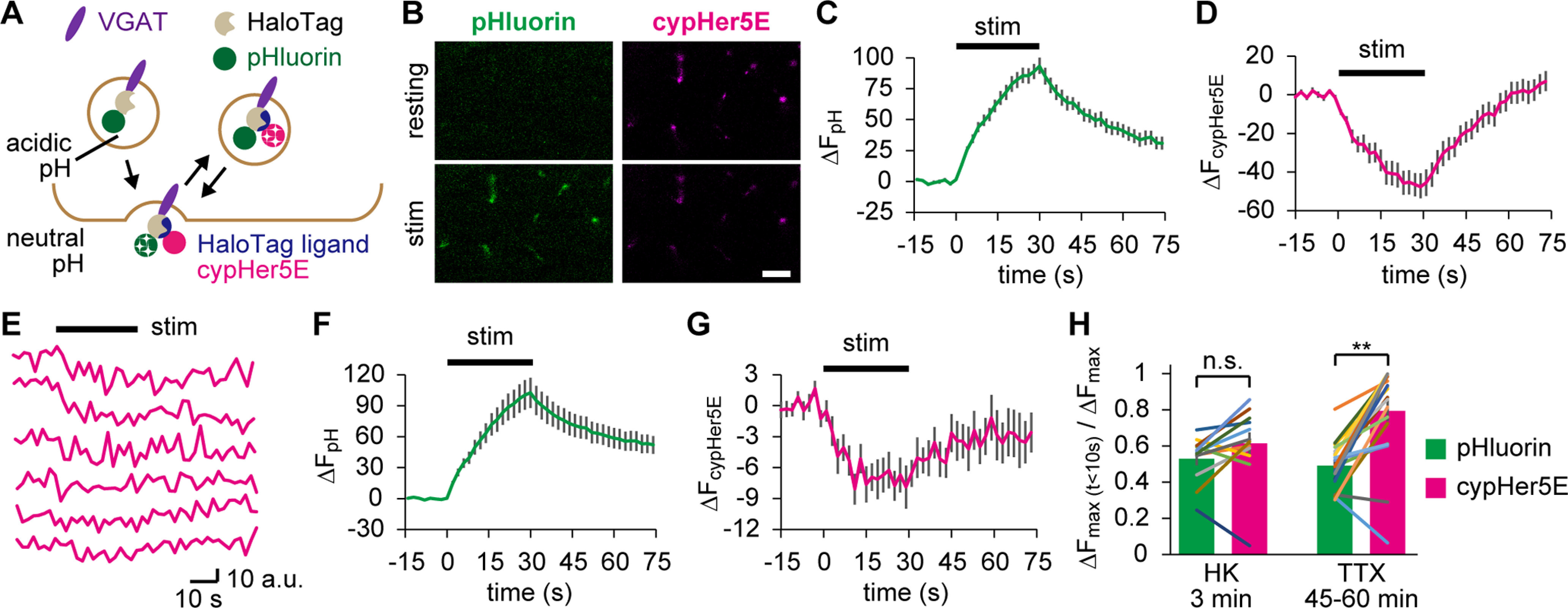
Spontaneously recycled SVs behaved like RRP vesicles in subsequent APs. A, Diagram represents HaloTag labeling with HL-cypHer5E, which allows SV imaging after the initial exocytosis. B, Confocal live images of pHluorin and cypHer5E at the NMJs before (resting) and after (stim) APs (20 Hz for 30 s). HL-cypHer5E was loaded by 3 min HK depolarization preceding the electrical stimulation. Scale bar, 10 μm. C, D, pHluorin fluorescence (C) and cypHer5E fluorescence (D) in response to APs (20 Hz for 30 s) measured at the NMJs prelabeled with HL-cypHer5E through 3 min HK depolarization (n = 12 experiments from 7 fish). The cypHer5E fluorescence, which is maximum at acidic pH, is not photostable; thus, its photobleaching was corrected (see Materials and Methods). E, cypHer5E fluorescence in response to APs (20 Hz for 30 s), where HL-cypHer5E was preloaded during 45-60 min incubation in TTX. Each trace is from an individual experiment. F, G, pHluorin fluorescence (F) and cypHer5E fluorescence (G) in response to APs (20 Hz for 30 s) measured at the NMJs preloaded with HL-cypHer5E during 45-60 min incubation in TTX (n = 20 experiments from 16 fish). H, The fraction of SVs exocytosed in the first 10 s of electrical stimulation was calculated for preloaded SVs (cypHer5E) and the total SVs (pHluorin). Preloading was performed either by HK stimulation or incubation in TTX. SVs preloaded in TTX fused significantly faster than that in total SVs (TTX 45-60 min; **p < 0.01, paired t test), which was not the case in SVs preloaded by HK (HK 3 min; p = 0.08, paired t test). Error bars indicate ±SEM.

Acknowledgments
This image is the copyrighted work of the attributed author or publisher, and ZFIN has permission only to display this image to its users. Additional permissions should be obtained from the applicable author or publisher of the image. Full text @ J. Neurosci.