IMAGE

Fig. 1

ID
ZDB-IMAGE-220505-10
Source
Figures for Dzementsei et al., 2022
Image
Figure Caption

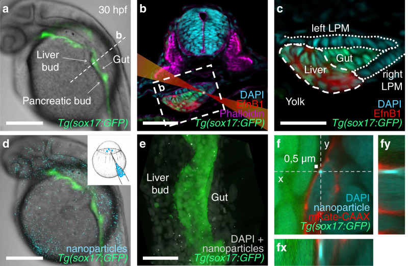
Fig. 1 Injected nanoparticles distribute throughout the embryo, including the foregut region in 28–30 hpf zebrafish embryos.

a Transgenic tg(-0.5 sox17:GFP)zf99 visualizes the endoderm in a living zebrafish embryo, including the foregut organ primordia. b Transverse section through the foregut region (dashed line in a) with schematized laser beam path (red) and bead (white). The foregut endoderm expresses transgenic sox17:GFP (green) and liver progenitors EphrinB1 (red); DAPI marks nuclei (cyan); and Phalloidin marks F-actin (magenta). c Magnification of the foregut region shown in (b) (white dashed rectangle). d 0.5 µm polystyrene particles (cyan) microinjected at the one-cell stage distribute throughout the embryo, including the endoderm (green), without causing apparent morphological defects. e Maximum intensity projection of confocal z-stack showing nanoparticle distribution (white) in the foregut region, GFP marks the endoderm. f Confocal image of a representative nanoparticle (light blue) located in the cytoplasm between the nucleus (darker blue) and plasma membrane (red) in a fixed embryo. (fy) and (fx) show orthogonal views in the “yz” plane (dashed line “y” in (f)) and “xz” plane (dashed line “x” in (f)), respectively. Scale bars a, d: 250 µm; b, e: 50 µm; c: 30 µm; f: 0,5 µm (scale bar next to bead).

Acknowledgments
This image is the copyrighted work of the attributed author or publisher, and ZFIN has permission only to display this image to its users. Additional permissions should be obtained from the applicable author or publisher of the image. Full text @ Commun Biol