
IMAGE
Figure 7—figure supplement 5.
- ID
- ZDB-IMAGE-220426-60
- Publication
- Cornean et al., 2022 - Precise in vivo functional analysis of DNA variants with base editing using ACEofBASEs target prediction
- All Figures
- Figures for Cornean et al., 2022
Image
|
Figure Caption
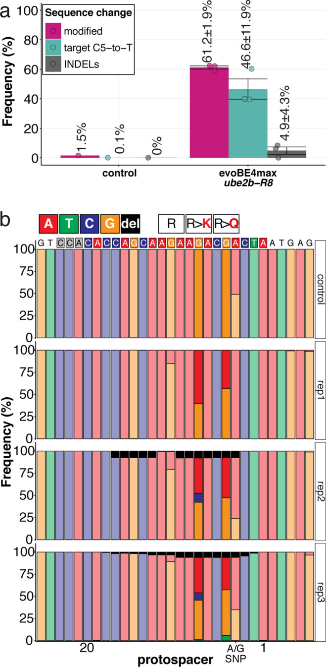
Figure 7—figure supplement 5.
The full complement of analyzed CVD associated phenotypes in cytosine base editing of
Acknowledgments
This image is the copyrighted work of the attributed author or publisher, and
ZFIN has permission only to display this image to its users.
Additional permissions should be obtained from the applicable author or publisher of the image.
Full text @ Elife