
|
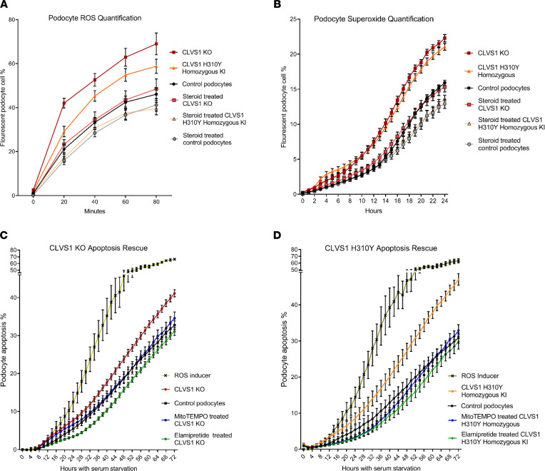
Figure 7
(A and B) Automated live-cell imaging and quantification of ROS levels in CLVS1-KO podocytes as well as controls using a fluorescent reporter of multiple ROS types, including hydrogen peroxide, peroxynitrite, and hydroxl radicals (A) (n > 20 for each group, P < 0.0001 for all time points for KO, P < 0.05 for all time points, and after 20 minutes for KI, 2-way ANOVA), as well as a second independent fluorescent reporter that detects superoxide generation (B) (n > 20 for each group, P < 0.001 for all time points after 10 hours, 2-way ANOVA), revealed an increase in ROS and superoxide accumulation in CLVS1-KO and homozygous KI podocytes that could be rescued with pretreatment with 1 μM dexamethasone. (C and D) The increased susceptibility to apoptosis in CLVS1-KO and homozygous KI podocytes (P < 0.05 for all time points after 36 hours) could be rescued by treatment with a superoxide scavenger or an ROS inhibitor (5 nM MitoTEMPO and 500 nM elamipretide) (n > 10 for all conditions, 2-way ANOVA). The ROS inducer pyocyanin (300 μM) and ROS inhibitor N-acetyl-l-cysteine (5 mM) were used as positive and negative controls, respectively, for all experiments. Error bars depict SEM.