IMAGE

FIGURE 5

ID
ZDB-IMAGE-220420-41
Source
Figures for Martins et al., 2022
Image
Figure Caption

FIGURE 5

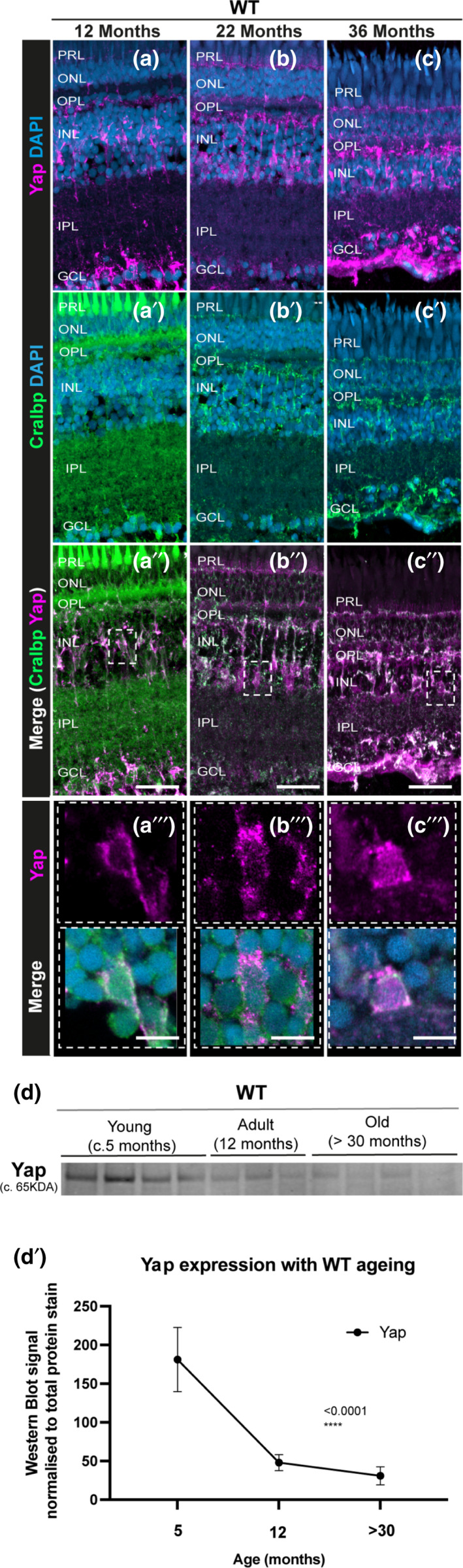
Yap expression is observed in the retinal MG throughout the zebrafish lifespan, but overall levels decrease with ageing. (a–c) The central retina immunolabelled for Yap expression (in magenta) and (a’–c’) Cralbp (MG cells, in green) in WT zebrafish at different ages (c. 12 months, 22 months, and 36 months). (a’’–c’’) Merge of DAPI, Yap and Cralbp staining and (a’’’–c’’’) respective inset showing Yap expression in MG cells at all stages observed. Representative images shown from N = 3 animals per age. Scale bars: 20 μm and 3 μm (in insets). (d–d’) Western blot of whole eyes and respective quantification show decreased Yap expression with ageing in WT zebrafish. N = 3–4

Acknowledgments
This image is the copyrighted work of the attributed author or publisher, and ZFIN has permission only to display this image to its users. Additional permissions should be obtained from the applicable author or publisher of the image. Full text @ Aging Cell