
|
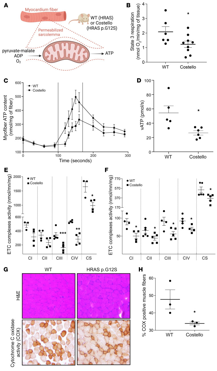
Figure 2 Mitochondrial bioenergetics is altered in situ in the CS mouse heart and skeletal muscle. (A) Permeabilized heart muscle fibers bioenergetics evaluation methods. (B) Rate of coupled (ADP-stimulated “state 3”) respiration was determined in situ using high-resolution respirometry (WT, n = 6; Costello, n = 9). (C and D) Rate of mitochondrial ATP synthesis (vATp) determined in heart-permeabilized muscle fibers (WT, n = 5; Costello, n = 6). (E) Electron transport chain (ETC) complex enzymatic activities determined in WT or Costello mouse model hearts (WT, n = 4; Costello, n = 4). (F) Respiratory chain complex enzymatic activities determined in WT or Costello mouse model skeletal muscle (WT, n = 4; Costello, n = 4). (G) Histo-enzymology staining of the respiratory chain complex IV (COX) specific activity. Muscle fibers and their nuclei were stained using H&E. Original magnification, ×1000. (H) Quantification of the COX-positive muscle fibers (WT, n = 3; Costello, n = 3). Data are expressed as the mean ± SEM. *P < 0.05, **P < 0.01, ***P < 0.001 (unpaired t test).