
Figure 3
|
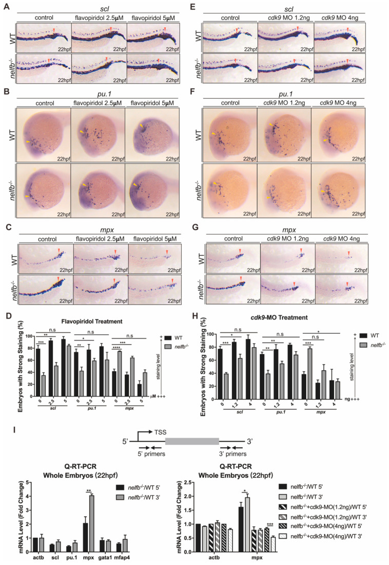
Figure 3 Inhibition of Pol II elongation rescues primitive hematopoiesis in nelfb−/− embryos. (A–C) WISH for scl (A), pu.1 (B) or mpx (C) at 22 hpf in WT and nelfb−/− embryos treated with DMSO, 2.5 µM flavopiridol, or 5 µM flavopiridol. (D) Quantification of WISH results in (A–C) (n = 30–50 embryos per group). (E–G) WISH for scl (E), pu.1 (F), or mpx (G) at 22 hpf in WT and nelfb−/− embryos without or with cdk9 MO injection (1.2 ng or 4 ng). (H) Quantification of WISH results in (E–G) (n = 30–50 embryos per group). (I) Upper panel shows the position of primers used in Q-RT-PCR analysis. Primers for the 5′ transcripts are located within 120 bp from transcription start site (TSS), and primers for the 3′ transcripts are in the 3′ coding region or 3′UTR. Lower-left panel shows Q-RT-PCR analysis of 5′ and 3′ transcripts of hematopoiesis-related genes in WT and nelfb−/− embryos at 22 hpf. Lower-right panel shows Q-RT-PCR analysis of 5′ and 3′ transcripts of mpx gene in 22 hpf nelfb−/− embryos without or with cdk9 MO injection (1.2 ng or 4 ng). Gene expression is normalized to the 5′ transcript of β-actin and shown as fold-change relative to WT, following the methods in the previous study [35]. All results are presented as the mean ± SD from three independent experiments (t test, * for p < 0.05, ** for p < 0.01, *** for p < 0.001, **** for p < 0.0001). Yellow arrowheads and red arrowheads in (A–C) and (E–G), respectively, indicate ALPM and PLPM. “+++” and “+” in (D,H) respectively represent strong staining and weak staining. MO, morpholino.