IMAGE

Fig. 3

ID
ZDB-IMAGE-220404-3
Source
Figures for Haraoka et al., 2022
Image
Figure Caption

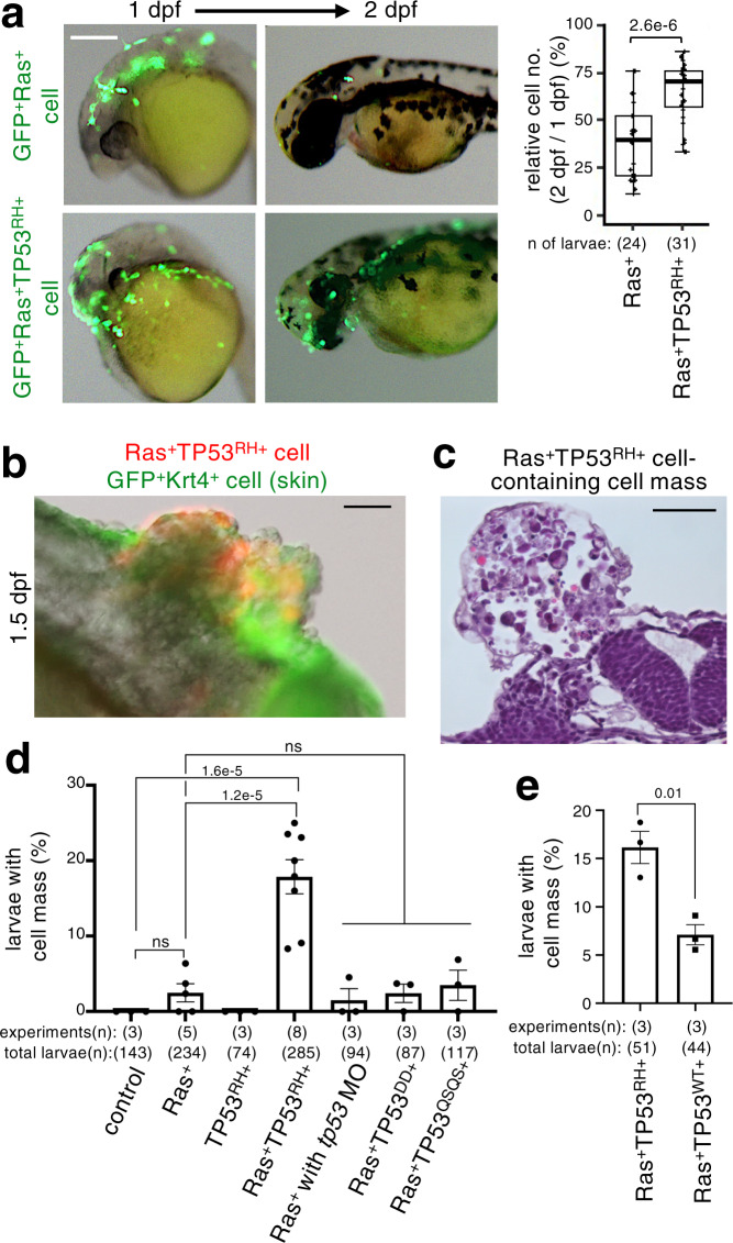
Fig. 3 TP53<sup>R175H</sup> mutation attenuates mosaic Ras<sup>G12V</sup> cell elimination and stimulates heterogeneous cell mass formation.

a Mosaic RasG12VTP53R175H cells remain in larval skin. Representative images show larvae with mosaically introduced GFP+RasG12V (GFP+Ras+) or GFP+RasG12VTP53R175H (GFP+Ras+TP53RH+) cells (green). Scale bar: 200 μm. Box plots of relative GFP+ cell number of 2 dpf/1 dpf ratio show first and third quartile, median is represented by a line, whiskers indicate the minimum and maximum. Each dot represents one larva. Unpaired two-tailed t-test was used. be TP53 gain of function, but not loss of function, stimulates heterogeneous cell mass formation. In b, representative images show cell mass in the trunk region of larvae with mosaically introduced mKO2+RasG12VTP53R175H (Ras+TP53RH+) cells (red) at 1.5 dpf. Skin surface layer cells were visualised with krt4p:gal4; UAS:EGFP (green). In c, representative image of haematoxylin and eosin staining of cell mass in the trunk region of larvae with mosaically introduced Ras+TP53+ cells. Scale bar: 50 μm. Graph in d and e shows the percentage of 1.5 dpf embryos with cell mass (Means ± SEM). Each dot represents an independent experimental result. In d, A two-tailed one-way ANOVA test with Sidak correction was used. In e, Unpaired two-tailed t-test was used. Note that cell mass formation was induced not only in the head region but also in the trunk and tail regions. Source data are provided as a Source Data file.

Figure Data
Acknowledgments
This image is the copyrighted work of the attributed author or publisher, and ZFIN has permission only to display this image to its users. Additional permissions should be obtained from the applicable author or publisher of the image. Full text @ Nat. Commun.