
|
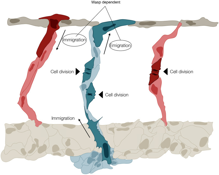
Fig. 7.
Illustration of the principal morphodynamic behaviours that differentially control regular vessel diameter formation in arteries and veins of the zebrafish trunk. A vISV (blue) is shown with two ECs undergoing division (black arrowhead), and with one cell immigrating from the PCV (the PCV vessel is not fully illustrated) and one cell emigrating to the DLAV (all morphodynamic events are highlighted in dark blue). Note that in the vISV, emigration and immigration are balanced, and cell numbers are increased by proliferation. Two arteries (red) are shown either side of the vISV. On the left, the aISV receives a cell by immigration from the DLAV (dark red). On the right, the aISV increases its cell number by cell division (dark red). In aISVs, these two processes are depicted as alternatives, as they seldom coincide. The directional movement of EC from vISV to the DLAV, and from the DLAV to aISV, requires WASp.