IMAGE

Figure 4

ID
ZDB-IMAGE-220324-8
Source
Figures for Hsu et al., 2022
Image
Figure Caption

Figure 4

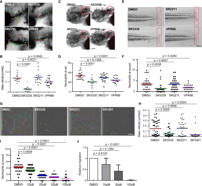
Pharmacological inhibition of Rora reduces neutrophil motility and chemotaxis in zebrafish and humans. (A, B) Tracks and quantification of neutrophil motility in zebrafish larva treated with SR3335 (100μM), SR2211 (100μM), or VPR66 (25μM). Scale bar, 200 µm. Three embryos, each from three different founders, were imaged. Quantification of neutrophils in one representative video is shown, Kruskal–Wallis test. (C, D) Representative images and quantification of neutrophils recruited to the infected ear in zebrafish larva treated with RORα specific inhibitor (SR3335, 100μM), RORγ specific inhibitor (SR2211, 100μM) or pan-ROR family inhibitor (VPR66, 25μM). Scale bar, 100 µm. (E, F) Representative images and quantification of neutrophils recruited to tail fin transection sites in zebrafish larva treated with SR3335 (100μM), SR2211 (100μM), or VPR66 (25μM). Scale bar: 500 µm. (C–F) The result from one representative experiment is shown as mean, Mann–Whitney test. (G, H) Representative tracks and mean velocity of primary human neutrophils treated with SR3335 (50μM), SR2211 (50μM), or SR1001 (pan-ROR family inhibitor) (50 µM) migrating towards fMLP in 3D matrigel. Scale bar, 100 µm. Representative results for three individual trials are shown. The result is presented as mean, Mann–Whitney test. (I) Neutrophil recruitments after zebrafish tail wounding at different dosages of SR3335 treatment compared to 1% DMSO treatment, Kruskal–Wallis test. (J) Transwell migration of primary human neutrophils treated with DMSO (0.1%) or SR3335 at 10, 30, or 100 μM toward 100 nM fMLP. Results are presented as mean ± s.d., from three independent experiments and normalized to DMSO (0.1%), Kruskal–Wallis test.

Acknowledgments
This image is the copyrighted work of the attributed author or publisher, and ZFIN has permission only to display this image to its users. Additional permissions should be obtained from the applicable author or publisher of the image. Full text @ Front Immunol