IMAGE

Figure 2

ID
ZDB-IMAGE-220224-2
Source
Figures for Wang et al., 2022
Image
Figure Caption

Figure 2

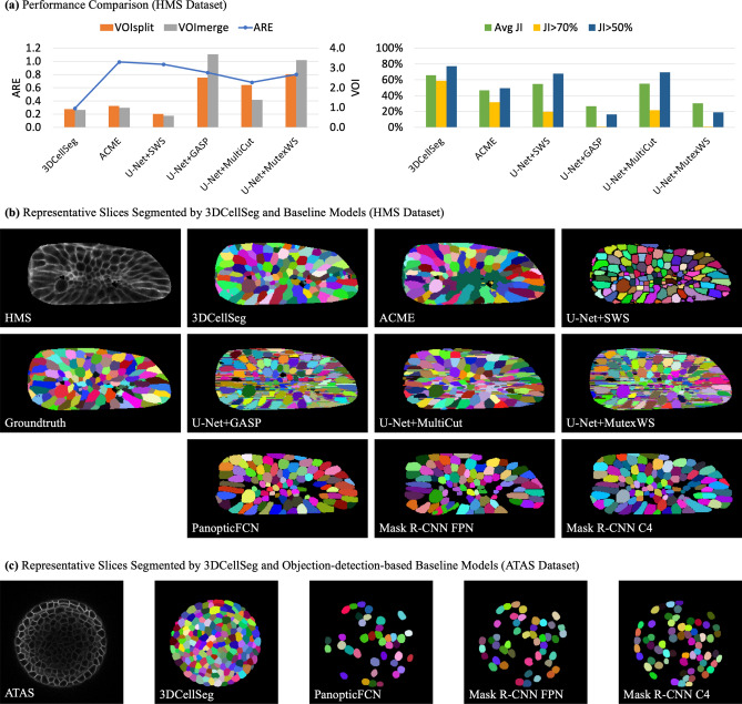
Model comparison and representative slices. [Note (a) shows the accuracies of different cell segmentation models for the HMS dataset. 3DCellSeg achieves the second best accuracy in ARE, VOIsplit, and VOImerge, and achieves the best accuracy in Avg JI, JI > 70%, and JI > 50% (the plots for DSC-related metrics are of high similarity to JI-related metrics). (b) and (c) show representative slices of different model segments. ACME tends to under-segment (see the dark green region which mis-classifies different cells as one cell) while U-Net + SWS tends to over-segment (see the over-segmented small cells in the central region). PanopticFCN, Mask R-CNN FPN, and Mask R-CNN C4 are accurate on the HMS dataset but they are severely under-segment on the ATAS dataset. The cellular images in (b) and (c) were generated by Python Matplotlib (https://matplotlib.org) using the HMS and ATAS49 datasets].

Acknowledgments
This image is the copyrighted work of the attributed author or publisher, and ZFIN has permission only to display this image to its users. Additional permissions should be obtained from the applicable author or publisher of the image. Full text @ Sci. Rep.