
|
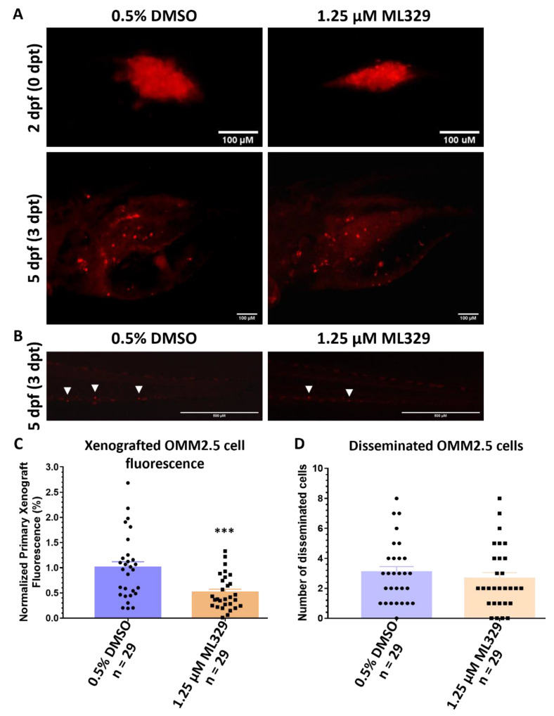
Figure 10
ML329 demonstrates anti-UM properties in zebrafish OMM2.5 xenografts. (A): Top panel shows OMM2.5 Dil-labeled cells xenografted into the perivitelline space of 2-day-old zebrafish larvae. Bottom panel presents zebrafish larvae 3 days post treatment (dpt) with 0.5% DMSO (n = 29) or 1.25 μM ML329 (n = 29). Days post fertilization (dpf). (B). Representative image of OMM2.5 Dil-labeled cells disseminated (white arrowhead) to the caudal vein plexus of zebrafish larvae at 3 dpt. (C): A significant (***, p = 0.0006) regression of the average normalized primary xenograft fluorescence of OMM2.5 Dil-labeled cells was observed when treated with 1.25 μM ML329. (D): No difference was detected in the average number of disseminated OMM2.5 Dil cells following treatment with 1.25 μM ML329 compared to vehicle control. Student’s T test was used for statistical analysis, with error bars presenting mean ± SEM.