
|
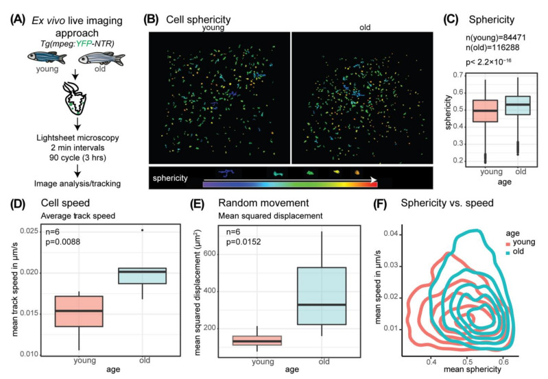
Figure 5
Macrophage morphology and behavior changes with age. (A) Schematic overview of the ex vivo live imaging approach using a reporter line expressing YFP under the control of the mpeg1.1 promoter (B) Sphericity of segmented and tracked objects (mpeg+ cells) at timepoint (tp) = 1 is color coded with red as high sphericity (round) and purple/blue as low sphericity. Age young: 12 months, age old: 4 years 7 months. The legend includes a color code for sphericity and representative example cell shapes. (C) Comparison of sphericity of all tracked objects (mpeg+ cells) at all time points analyzed in old and young fish hearts reveals a significant shift to higher average cell sphericity in the old. Welch two-sample t-test p < 2.2 × 10⁻¹⁶, age young: 3 × 6 months and 3 × 12 months, age old: 3 × 4 years 7 months and 3 × 5 years. (D) The boxplot illustrates the mean track speed of all segmented tracks analyzed per biological replicate. 6 old and 6 young hearts were analyzed. Quantification of average speed of mpeg+ cells in old and young fish ventricles indicate a faster cell movement in old. n = 6; two-sample t-test p = 0.0088, age young: 3 × 6 months and 3 × 12 months, age old: 3 × 4 years 7 months and 3 × 5 years. (E) The boxplot illustrates the average mean squared displacement analyzed per heart of 6 old and 6 young hearts. Quantification of mean squared displacement of mpeg+ cells in old and young ventricles reveals a higher random movement in old. n = 6, Wilcoxon rank sum test p = 0.0152, age young: 3 × 6 months and 3 × 12 months, age old: 3 × 4 years 7 months and 3 × 5 years. (F) Density plot showing the average cell sphericity per track vs. average speed per track reveals a shift of cell populations in old towards more roundish and to faster cells.