
|
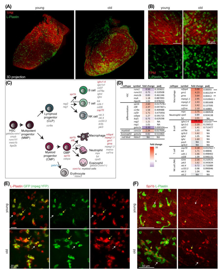
Figure 4
Immune cells accumulate in ventricles upon aging. (A) Whole-mount immunostaining of old (4 years) and young (1 year) fish hearts for the pan-leukocyte marker L-Plastin (green) and the muscle marker Tropomyosin (TPM) (red) are shown. Lightsheet microscopy and 3D projection reveal increased cell numbers and cell cluster formation in old hearts. (B) Higher magnification images of old and young hearts stained with L-Plastin (green) and TPM (red) reveals that immune cell numbers increase and cell morphology changes to be more roundish in old hearts. Scale bar: 50 µm. Whole-mount immunostaining and lightsheet microscopy. (C) Scheme of different immune cell types and their respective lineages in zebrafish. Known marker genes for different cell populations as well as for differentiation into specialized cell types are listed. Genes with significantly up or down regulated expression in old are highlighted in red or blue, respectively. (D) Immune cell markers (from (C)), their respective fold change with age and adjusted p-value (padj) are listed (transcriptional profiling from Figure 1, n = 3). Fold change values are color-coded with up in red, no change white and down in blue. NA: not available. *: padj < 0.05; **: padj < 0.01, ***: padj < 0.001 (E) Whole-mount immunostaining of young (1 year) and old (3 years) fish hearts of a mpeg:YFP reporter line for the pan-leukocyte marker L-Plastin (red) and GFP (green) reveals that a large subset of L-Plastin+ cells are mpeg-positive. White arrow indicates a single positive immune cell and yellow arrow indicates a double positive cell. Lightsheet microscopy. Scale bars: 20 µm. (F) Whole-mount immunostaining of young (1 year) and old (4 years) fish hearts for the pan-leukocyte marker L-Plastin (green) and Spi1b (red) reveal large subset of L-Plastin cells are Spi1b-positive. White arrow indicates a single positive immune cell and yellow arrow indicates a double positive cell. Lightsheet microscopy. Scale bars: 50 µm.